Selezione di pubblicazioni scientifiche del gruppo di ricerca di IMAGO7 e del FiRMLab
Per una lista completa, consultare le pagine Google Scholar dei ricercatori →
2026
Ambrosi P, Lancione M, Cecchi P, Tosetti M, Biagi L
Robustness, spatial detail, and pitfalls of fixed ICA dimensionality in resting-state fMRI networks at 1.5, 3, and 7 T
Frontiers in Neuroscience, 19:1731143, 2026
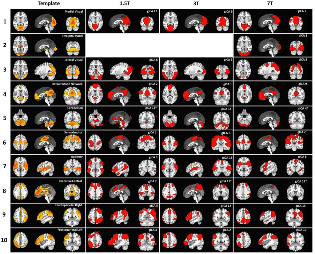
ABSTRACT: Resting-state fMRI functional connectivity analysis is usually performed with seed-based methods that strongly rely on user-dependent definitions of regions of interest. Data-driven methods like independent component analysis (ICA) can mitigate this need. However, the number of components that should be expected in an fMRI acquisition, which determines the model order of the ICA, is not defined, and it is not uniformly chosen across studies. This variability is further complicated by the dependence of component number on field strength, with higher field strengths typically yielding more detectable components. Therefore, relying on a predetermined number may influence the results. Here, we compare functional maps obtained through ICA analysis at different magnetic field strengths and at various levels of spatial detail. Our results confirm the presence of the most frequently reported resting-state networks across field strengths and demonstrate that higher magnetic field strength enables more robust detection of functional networks with greater spatial detail. We also show that: (1) fixing the number of components, although improving interpretability of group results, may provide an incomplete picture of brain function; (2) a greater number of components is consistently identified at higher field strength, suggesting that the model order should be adapted according to both field strength and spatial detail.
2025
Acquafredda M, Kurzawski JW, Biagi L, Tosetti M, Morrone MC, Binda P
The pulvinar regulates plasticity in human visual cortex
Science advances, 11:48, eadw9988, 2025
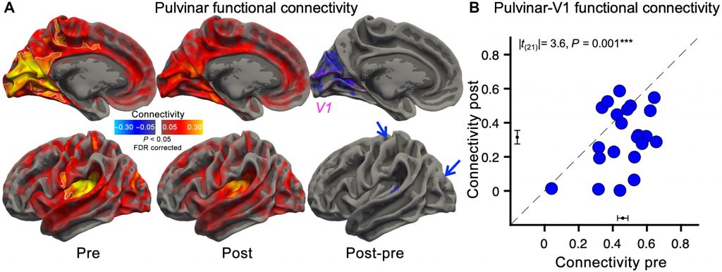
ABSTRACT: In normally sighted human adults, 2 hours of monocular deprivation is sufficient to transiently alter ocular dominance. Here, we show that this is associated with a reduction of functional connectivity between the pulvinar and primary visual cortex (V1), selective for the pulvinar-to-V1 directionality. Across participants, the strength of the pulvinar-to-V1 connectivity was negatively correlated with the ocular dominance shift, implying less plasticity in participants with stronger influence of the pulvinar over V1. Our results support a revised model of adult V1 plasticity, where short-term reorganization is gated by modulatory signals relayed by the pulvinar.
Marmin S, Arduino A, Cencini M, Lancione M, Biagi L, Tosetti M, Zilberti L
Linking dielectric dispersion and age in brain tissues via water content-based Electric Properties Tomography
NeuroImage, 322, 121559, 2025
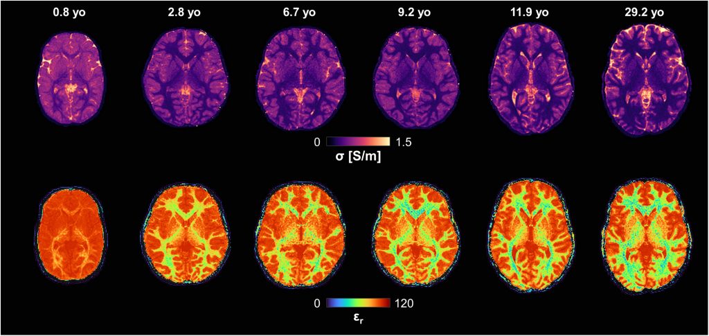
ABSTRACT: Popular dielectric dispersion models of biological tissues, which describe dielectric properties as a function of frequency, do not account for age-related variations. In particular, existing databases have limited validity in pediatric populations. In this work, we applied water content-based Electric Properties Tomography in vivo to healthy subjects across the lifespan to incorporate age-related information into dielectric dispersion models of white and grey matter. Water content, derived from magnetic resonance fingerprinting-based mapping, was modelled as a function of age. The age-water relationships was then integrated with Cole-Cole dispersion via water-dependent permittivity and conductivity equations. The resulting model allows obtaining age-specific conductivity and permittivity of brain tissues at frequencies higher than 50 MHz. In addition, it provides confidence intervals accounting for both intra-subject and inter-subject variability. In terms of applications for brain studies, this model enables age-specific electromagnetic simulations for pediatric subjects and evaluations on the safety of electromagnetic exposure in developing brains. An open code is freely available online to compute electrical properties and their uncertainties as a function of frequency and age.
Lancione M, Cencini M, Aquino D, Baldoli C, Elia M, Ghielmetti F, Montanaro D, Neri I, Nigri A, Pasquariello R, Pettinato S, Romano S, Sbrizzi A, Scifo P, van der Heide O, Versteeg E, Biagi L, Tosetti M
Repeatability and reproducibility of joint T1-T2 transient-state relaxometry across multiple vendors and implementations at 3T in phantom and human brain
NeuroImage, 320, 121471, 2025
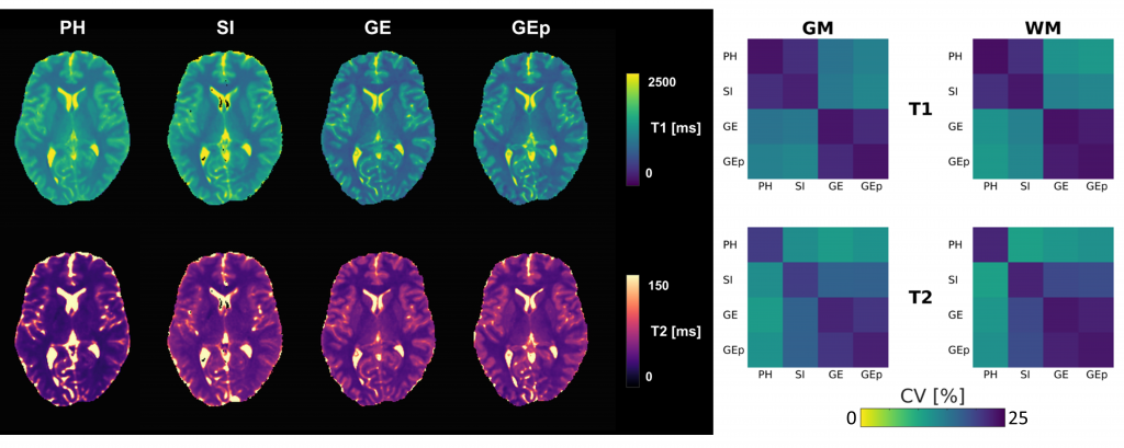
ABSTRACT: Transient-state relaxometry (TSR) enables rapid estimation of T1 and T2 relaxation times. To support its broader adoption in multi-center studies, it is essential to assess the consistency of its implementation across different MRI vendors.
This work aimed to assess accuracy, repeatability, and inter-vendor reproducibility of jointly measured T1 and T2 maps based on TSR at 3T. To achieve this goal, a phantom and five volunteers were scanned in a traveling-brain study at four 3T MRI systems from three manufacturers.
In the phantom study, Bland-Altman analysis and coefficients of variation (CV) were used to assess accuracy, and repeatability and reproducibility, respectively. Subsequently, in-vivo inter-site variability was evaluated via ANOVA and by computing voxelwise CVs and biases associated with sites were measured via a general linear model (GLM).
Excellent accuracy, repeatability, and reproducibility were obtained for the phantom. In-vivo, we found excellent repeatability (CV < 4.5%) and generally good inter-site and inter-vendor reproducibility, though significant variability was found across different TSR implementations. The GLM analysis revealed site-related biases of approximately 100 ms for T1 and 2 ms for T2 in solid brain tissues. These differences may be attributable to different magnetization transfer effects and residual B1+ inhomogeneities due to imperfect calibration.
Our findings demonstrate that the bias introduced by the use of different TSR implementations needs to be considered carefully in order to perform in-vivo multi-center studies.
Calzoni T, Donatelli G, Migaleddu G, Lancione M, Cecchi P, Biagi L, Caniglia M, Ceravolo R, Cosottini M
Hypertrophic Olivary Degeneration: a 7 Tesla Advanced Imaging Case Report
Frontiers in Neuroscience, 19:1656655, 2025
ABSTRACT: Introduction: A 50-years-old patient developed ataxia, nystagmus and palatal tremor. Conventional MRI revealed inferior olivary nuclei enlargement and hyperintensity in T2-weighted images, posing diagnosis of hypertrophic olivary degeneration (HOD). The patient’s past medical history reported proton-therapy for an VIII cranial-nerve Schwannoma. Here we aimed to investigate the potential alterations involving tracts and nuclei composing the dentato-rubro-olivary pathway (Guillain-Mollaret triangle) using an advanced ultra-high field (7T) MRI protocol.
Materials and Methods: The patient underwent a brain 7T-MRI exam including a multi-echo gradient-echo sequence for quantitative susceptibility mapping and diffusion tensor imaging (DTI). The DTI dataset was elaborated for tractography and computation of tensor metrics.
Results: 7T-MRI allowed the depiction of the brainstem tracts and nuclei composing the Guillain-Mollaret triangle. Both qualitative and quantitative analysis of these structures demonstrated damage to the right red nucleus and to the dentato-rubral tracts bilaterally. These findings are consistent with the pathophysiology of HOD and were confirmed in a follow-up MRI.
Discussion: This study highlights the capability of 7T-MRI to depict and investigate brainstem substructures such as tracts and nuclei. To the best of our knowledge, this is the first study to depict all tracts composing the Guillain-Mollaret triangle and directly document their alterations in HOD.
Curzio O, De Pasquale CF, Maestro S, Belmonti V, Biagi L, Tosetti M, Muratori F, Pasquariello R, Retico A, Calderoni S
Body Mass Index Impacts on Gray Matter Volume in Developmental Restrictive Anorexia Nervosa: A Voxel-Based Morphometry Study
Nutrients, 17(16):2620, 101062, 2025
ABSTRACT: Background/Objectives: Previous magnetic resonance imaging (MRI) investigations reported brain alterations in anorexia nervosa restricting type (AN-R); however, the number of existing structural neuroimaging studies in the developmental age is limited. Here, we analyzed the volumetric brain differences between adolescent patients with AN-R and control peers, and possible correlations between brain volumes and clinical features. Methods: The sample comprised 47 adolescent females with AN-R (mean age: 15.0 years, SD = 1.4) who underwent structural MRI within one month of admission to a tertiary care university hospital, and 39 typically developing controls matched for sex and age. The patients were clinically characterized by standardized interviews/questionnaires. Using the voxel-based morphometry (VBM) technique, possible significant volumetric brain differences between the two groups were analyzed. Moreover, correlations between altered brain regions and clinical (i.e., body mass index (BMI) and disease duration) or psychopathological variables were investigated. Results: An overall reduction in gray matter (GM) volume with a concomitant increase in cerebrospinal fluid (CSF) is observed in AN-R patients; these alterations correlate with a lower BMI. The reduction in GM volume affects the frontal and parietal regions involved in the cognitive processes that underlie and sustain the AN-R clinical features. Conclusions: These results add to the current knowledge of the AN-R pathophysiology and pave the way for the development of brain imaging biomarkers for AN in the developmental age.
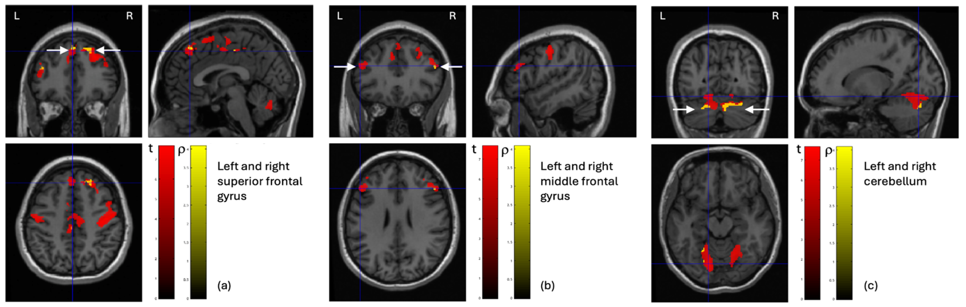
Desiato G, Bosco P, Cintoli S, Biagi L, Braschi C, Del Nero C, Minichiello I, Noale M, Faggiani E, Rossi A, Pozzi D, Kallikourdis M, Pratali L, Maggi S, Tognoni G, Berardi N, Maffei L, Sale A, Tosetti M, Matteoli M
An inflammatory fingerprint in mild cognitively impaired patients is reversed by physical and cognitive training
Brain, Behavior, & Immunity-Health, 101062, 2025
ABSTRACT: Background: Alzheimer’s disease (AD) is a major global health concern, with number of affected individuals expected to rise to 139 million by 2050. Lifestyle factors play a significant role in modulating cognitive decline, and multidomain interventions have demonstrated effectiveness in improving outcomes for populations at risk. The Train the Brain (TTB) program—a combined physical and cognitive training delivered in a social setting—has previously demonstrated cognitive benefits within 7 months. However, the underlying biological mechanisms remain unclear. Given the role of inflammation in aging and neurodegeneration, we investigated whether specific immune biomarkers reflect the efficacy of this intervention.
Methods: We enrolled 76 individuals with Mild Cognitive Impairment (MCI) aged 65–80, into the TTB program. Participants underwent neurological assessment, MRI and blood sampling at baseline and after the intervention. Plasma levels of a comprehensive panel of immune – related biomarkers were measured through Proquantum and ELLA platforms.
Results: At baseline, MCI participants displayed elevated levels of IL-17A, CX3CL1, CCL11, with a borderline increase of IL-6 and TNFα. Following the TTB intervention, we observed reductions in IL-6, IL-17A, TNFα, and CCL11 levels. In contrast, anti–inflammatory cytokines (IL-10, TGFβ, IL-4) and BDNF declined in control group but were maintained or increased in the intervention group.
Conclusion: The TTB intervention not only improved cognitive and physical outcomes but also modulated key immune markers associated with neuroinflammation and aging. IL-10, in particular, emerged as potential peripheral biomarker of training efficacy. These findings support the utility of immune profiling in monitoring response to multidomain interventions and guiding personalized strategies for cognitive risk reduction.
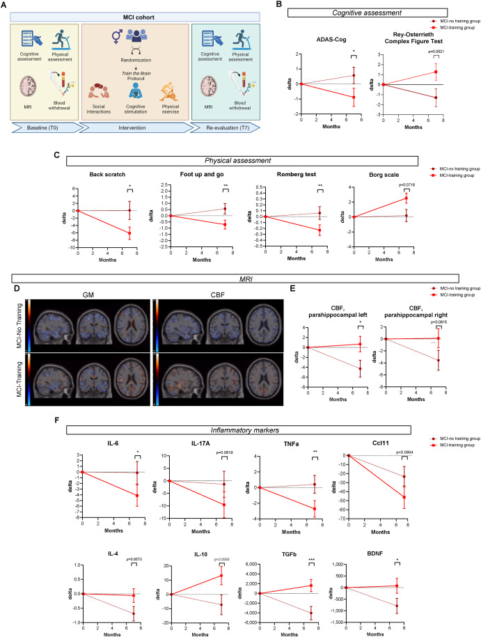
Lancione M, Donatelli G, Migaleddu G, Cencini M, Bosco P, Costagli M, Ceravolo R, Cosottini M, Tosetti M, Biagi L
High resolution multi-parametric probabilistic in vivo atlas of dorsolateral nigral hyperintensity via 7 T MRI
Scientific Data, 12:958, 2025
ABSTRACT: The role of Nigrosome 1 (N1) in neurodegeneration and motor disorders, particularly in Parkinson’s disease (PD), is increasingly recognized. The study of this region using quantitative measures, such as iron quantification through Quantitative Susceptibility Mapping (QSM), can provide enlightening insights into some pathological features of these diseases representing important biomarkers. However, the small size and the vanishing contrast with respect to the surrounding substantia nigra in PD patients make the segmentation of N1 challenging. For this reason, we provide a probabilistic atlas of the N1 portion corresponding to the swallow-tail hyperintensity, hereafter referred to as the Dorsolateral Nigral Hyperintensity (DNH), created on a high-resolution multi-parametric template from T1-weighted, T2*-weighted, and QSM images acquired in vivo at 7 T. The atlas also includes quantitative T2* and R2* templates and is provided in the MNI standard space. It aims to facilitate the study of N1, avoiding operator-dependent biases in segmentations, and allowing the standardisation of the quantitative assessment.
Cervelli R, Cencini M, Aringhieri G, Silvestrini B, Cacciato Insilla A, Campani D, Ghinolfi D, De Simone P, Tosetti M, Crocetti L
Ex-vivo 7T MRI of human explanted cirrhotic liver with HCC: quantitative and qualitative evaluation with radiological-pathological correlation
La radiologia medica, 130, 567–576, 2025
ABSTRACT: Introduction: Hepatocellular carcinoma’s (HCC) pathological grading is a recognized factor influencing intrahepatic recurrence after treatment. Thus, understanding the HCC heterogeneity is crucial to select the best treatment option aiming at personalized medicine. 7T MRI can provide qualitative and quantitative data, potentially identifying imaging biomarkers for lesions characterization.
Materials and methods: From May 2019 to December 2019, all explanted livers of patients undergoing liver transplant were enrolled. All patients underwent whole body CT before liver transplant and all the explanted livers were evaluated (ex-vivo) by 7T MRI within 12 h from liver removal with qualitative and quantitative acquisitions, including 2D/3D magnetic resonance fingerprinting (MRF). First, two expert radiologists qualitatively and quantitatively evaluated the imaging data focusing on both lesions and surrounding tissue, comparing conventional and MRF sequences. Then, specimens were evaluated by an expert pathologist regarding both liver tissues and lesions, particularly focusing on HCC grading.
Conclusions: This work may represent the first step supporting the introduction of quantitative MR imaging (including MRF) in the clinical practice. Along with conventional protocol and dynamic contrast enhancement, the integration of quantitative MR imaging can provide imaging biomarkers useful to identify HCC lesions more prone to recurrence, leading to a better patient selection, according to a personalized medicine approach.
2024
Hagberg GE, Golay X, Tosetti M
Towards quantitative MRI for the clinic
Journal of Neuroimaging, 124, 2024
PREAMBLE: Magnetic Resonance (MR) is an effective tool for clinical diagnosis, staging, monitoring, treatment planning, and assessing response to therapy. Measurements of anatomical, physiological, and biochemical characteristics of the organs through MR, referred to as quantitative imaging, is becoming increasingly used in clinical research for drug and medical device development and clinical decision-making.
Pathological conditions often cause alterations in tissues, whose intrinsic physical parameters can be measured by different MR techniques. The presence of such alterations on images has been extensively used as indications of disease; however, to classify alterations as biomarkers of disease, it is necessary to introduce quantitative measurements in the clinical setting. This in turn is placing a greater emphasis on clinical validation and planning techniques using MRI. The transition from standard (or qualitative) MRI to quantitative MRI is challenging and requires several actions to achieve interpretation of the pathological results with high accuracy, sensitivity, specificity, and predictive value. To increase the sensitivity of the measuring device to changes in quantitative imaging biomarkers associated with the disease, the entire process needs to be very clearly defined, including accurate definition of acquisition sequences, implementation of standardised methods of analysis, verification of reproducibility, as well as knowledge of variability from the scanner and other sources. As with any measurement, a quantitative MRI assessment should not only provide a number, but also its associated uncertainty. As for comparison with normal values, ranges and variabilities should be provided to enable proper comparison. This approach is crucial in the era of personalised medicine and when such quantitative measurements are used as outcome measures in multi-centre studies or clinical trials.
As MRI is steadily turning quantitative, thus novel possibilities based on innovative biomarkers are being developed and come within reach of routine clinical practice.
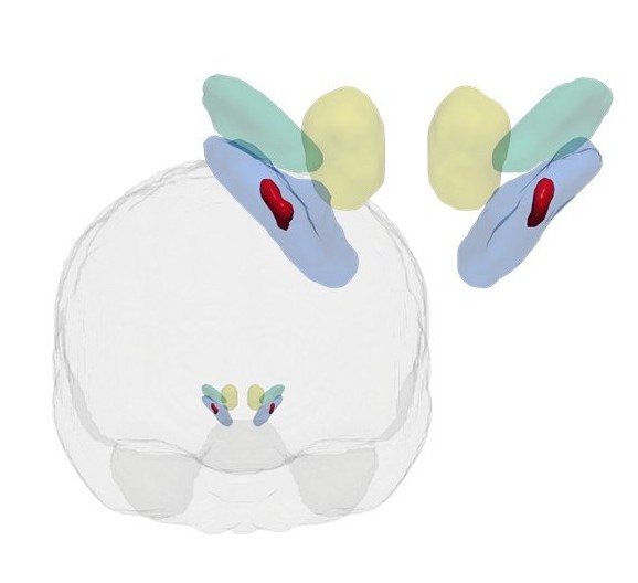
Cosottini M, Calzoni T, Lazzarotti GA, Grigolini A, Bosco P, Cecchi P, Tosetti M, Biagi L, Donatelli G
Time-of-flight MRA of intracranial vessels at 7 T
European Radiology Experimental, 8:68, 2024
ABSTRACT: Background: Three-dimensional time-of-flight magnetic resonance angiography (TOF-MRA) is a largely adopted non-invasive technique for assessing cerebrovascular diseases. We aimed to optimize the 7-T TOF-MRA acquisition protocol, confirm that it outperforms conventional 3-T TOF-MRA, and compare 7-T TOF-MRA with digital subtraction angiography (DSA) in patients with different vascular pathologies.
Methods: Seven-tesla TOF-MRA sequences with different spatial resolutions acquired in four healthy subjects were compared with 3-T TOF-MRA for signal-to-noise and contrast-to-noise ratios as well as using a qualitative scale for vessel visibility and the quantitative Canny algorithm. Four patients with cerebrovascular disease (primary arteritis of the central nervous system, saccular aneurism, arteriovenous malformation, and dural arteriovenous fistula) underwent optimized 7-T TOF-MRA and DSA as reference. Images were compared visually and using the complex-wavelet structural similarity index.
Results: Contrast-to-noise ratio was higher at 7 T (4.5 ± 0.8 (mean ± standard deviation)) than at 3 T (2.7 ± 0.9). The mean quality score for all intracranial vessels was higher at 7 T (2.89) than at 3 T (2.28). Angiogram quality demonstrated a better vessel border detection at 7 T than at 3 T (44,166 versus 28,720 pixels). Of 32 parameters used for diagnosing cerebrovascular diseases on DSA, 27 (84%) were detected on 7-T TOF-MRA; the similarity index ranged from 0.52 (dural arteriovenous fistula) to 0.90 (saccular aneurysm).
Conclusions: Seven-tesla TOF-MRA outperformed conventional 3-T TOF-MRA in evaluating intracranial vessels and exhibited an excellent image quality when compared to DSA. Seven-tesla TOF-MRA might improve the non-invasive diagnostic approach to several cerebrovascular diseases.
Donatelli G, Migaleddu G, Cencini M, Cecchi P, D’Amelio C, Peretti L, Buonincontri G, Tosetti M, Costagli M, Cosottini M
Detection of pathological contrast enhancement with synthetic brain imaging from quantitative multiparametric MRI
Journal of Neuroimaging, 34:4, 2024
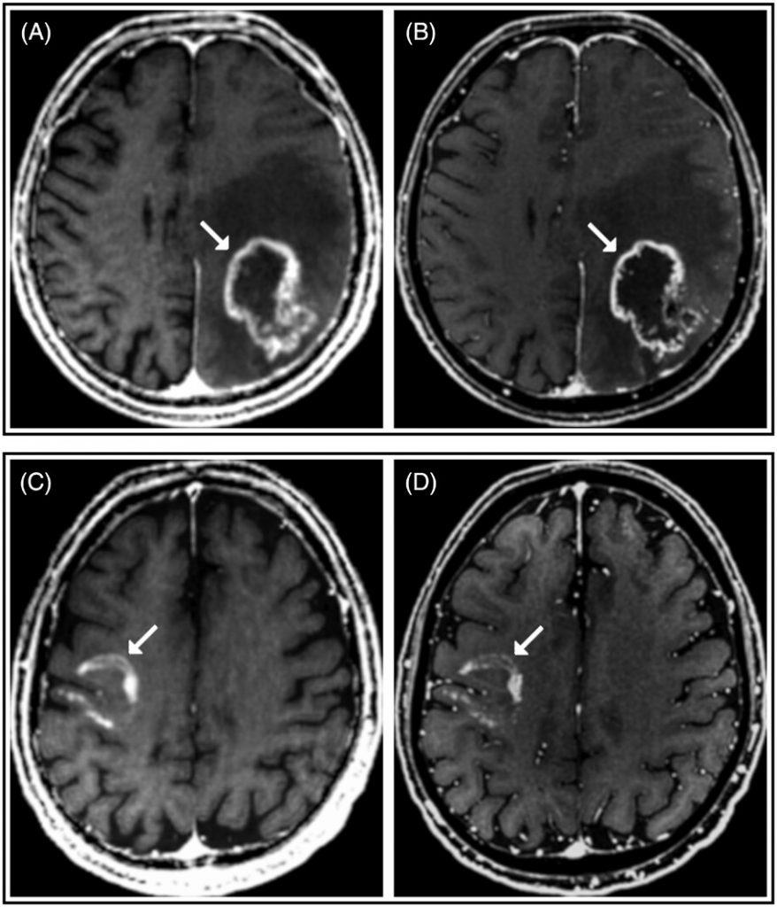
ABSTRACT: Background and Purpose: We aimed to test whether synthetic T1-weighted imaging derived from a post-contrast Quantitative Transient-state Imaging (QTI) acquisition enabled revealing pathological contrast enhancement in intracranial lesions.
Methods: The analysis included 141 patients who underwent a 3 Tesla-MRI brain exam with intravenous contrast media administration, with the post-contrast acquisition protocol comprising a three-dimensional fast spoiled gradient echo (FSPGR) sequence and a QTI acquisition. Synthetic T1-weighted images were generated from QTI-derived quantitative maps of relaxation times and proton density. Two neuroradiologists assessed synthetic and conventional post-contrast T1-weighted images for the presence and pattern of pathological contrast enhancement in intracranial lesions. Enhancement volumes were quantitatively compared.
Results: Using conventional imaging as a reference, synthetic T1-weighted imaging was 93% sensitive in revealing the presence of contrast enhancing lesions. The agreement for the presence/absence of contrast enhancement was almost perfect both between readers (k = 1 for both conventional and synthetic imaging) and between sequences (k = 0.98 for both readers). In 91% of lesions, synthetic T1-weighted imaging showed the same pattern of contrast enhancement visible in conventional imaging. Differences in enhancement pattern in the remaining lesions can be due to the lower spatial resolution and the longer acquisition delay from contrast media administration of QTI compared to FSPGR. Overall, enhancement volumes appeared larger in synthetic imaging.
Conclusions: QTI-derived post-contrast synthetic T1-weighted imaging captures pathological contrast enhancement in most intracranial enhancing lesions. Further comparative studies employing quantitative imaging with higher spatial resolution is needed to support our data and explore possible future applications in clinical trials.
Lancione M, Cencini M, Scaffei E, Cipriano E, Buonincontri G, Schulte RF, Pirkl CM, Buchignani B, Pasquariello R, Canapicchi R, Battini R, Biagi L, Tosetti M
Magnetic resonance fingerprinting-based myelin water fraction mapping for the assessment of white matter maturation and integrity in typical development and leukodystrophies
NMR in Biomedicine, e5114, 2024
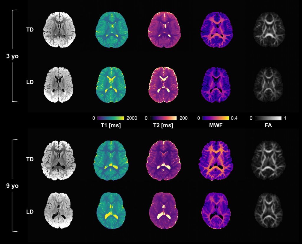
ABSTRACT: A quantitative biomarker for myelination, such as myelin water fraction (MWF), would boost the understanding of normative and pathological neurodevelopment, improving patients’ diagnosis and follow-up. We quantified the fraction of a rapidly relaxing pool identified as MW using multicomponent three-dimensional (3D) magnetic resonance fingerprinting (MRF) to evaluate white matter (WM) maturation in typically developing (TD) children and alterations in leukodystrophies (LDs). We acquired DTI and 3D MRF-based R1, R2 and MWF data of 15 TD children and 17 LD patients (9 months–12.5 years old) at 1.5 T. We computed normative maturation curves in corpus callosum and corona radiata and performed WM tract profile analysis, comparing MWF with R1, R2 and fractional anisotropy (FA). Normative maturation curves demonstrated a steep increase for all tissue parameters in the first 3 years of age, followed by slower growth for MWF while R1, R2 and FA reached a plateau. Unlike FA, MWF values were similar for regions of interest (ROIs) with different degrees of axonal packing, suggesting independence from fiber bundle macro-organization and higher myelin specificity. Tract profile analysis indicated a specific spatial pattern of myelination in the major fiber bundles, consistent across subjects. LD were better distinguished from TD by MWF rather than FA, showing reduced MWF with respect to age-matched controls in both ROI-based and tract analysis. In conclusion, MRF-based MWF provides myelin-specific WM maturation curves and is sensitive to alteration due to LDs, suggesting its potential as a biomarker for WM disorders. As MRF allows fast simultaneous acquisition of relaxometry and MWF, it can represent a valuable diagnostic tool to study and follow up developmental WM disorders in children.
Guan X, Lancione M, Ayton S, Dusek P, Langkammer C, Zhang M
NeuroImaging of Parkinson’s disease by Quantitative Susceptibility Mapping
NeuroImage, 120547, 2024
ABSTRACT: Parkinson’s disease (PD) is a common neurodegenerative disease, and apart from a few rare genetic causes, its pathogenesis remains largely unclear. Recent scientific interest has been captured by the involvement of iron biochemistry and the disruption of iron homeostasis, particularly within the brain regions specifically affected in PD. The advent of Quantitative Susceptibility Mapping (QSM) has enabled non-invasive quantification of brain iron in vivo by MRI, which has contributed to the understanding of iron-associated pathogenesis and has the potential for the development of iron-based biomarkers in PD. This review elucidates the biochemical underpinnings of brain iron accumulation, details advancements in iron-sensitive MRI technologies, and discusses the role of QSM as a biomarker of iron deposition in PD. Despite considerable progress, several challenges impede its clinical application after a decade of QSM studies. The initiation of multi-site research is warranted for developing robust, interpretable, and disease-specific biomarkers for monitoring PD disease progression.
Celardo G, Scaffei E, Buchignani B, Donatelli G, Costagli M, Cristofani P, Canapicchi R, Pasquariello R, Tosetti M, Battini R, Biagi L
Case report: Exploring chemoradiotherapy-induced leukoencephalopathy with 7T imaging and quantitative susceptibility mapping
Frontiers in Neurology, 15: 1362704, 2024
ABSTRACT: Chemotherapy and radiotherapy are widely used in the treatment of central nervous system tumors and acute lymphocytic leukemia even in the pediatric population. However, such treatments run the risk of a broad spectrum of cognitive and neurological deficits. Even though the correlation with cognitive decline is still not clear, neuroradiological defects linked to white matter injury and vasculopathies may be identified. Thanks to the use of 7T MRI it is possible to better define the vascular pattern of the brain lesions with the added advantage of identifying their characteristics and anatomical localization, which, however, are not evident with a conventional brain scan. Moreover, the use of Quantitative Susceptibility Mapping (QSM) makes it possible to discriminate between calcium deposits on vessels (chemo-radiation-induced) and hemoglobin deposition in radio-induced cavernomas, speculating, as a result, about the pathophysiology of iatrogenic brain damage. We describe the case of a 9 year-old boy with a T-type acute lymphoid leukemia who had previously been treated with polychemotherapy and high-dose RT. To better define the child’s neuroradiological pattern, 7T MRI and QSM were performed in addition to conventional imaging examinations. Our case report suggests the potential usefulness of a QSM study to distinguish radio-induced vascular malformations from mineralizing microangiopathy.
2023
Donatelli G, Emmi A, Costagli M, Cecchi P, Macchi V, Biagi L, Lancione M, Tosetti M, Porzionato A, De Caro R, Cosottini M
Exploring brainstem anatomy with 7T-MRI: in vivo assessment and ex vivo comparison
European Radiology Experimental, 7:71, 2023
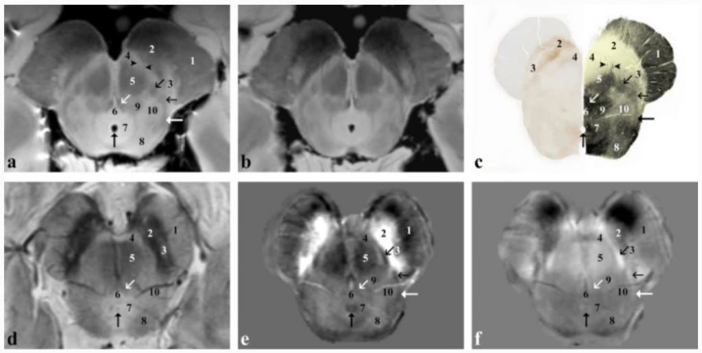
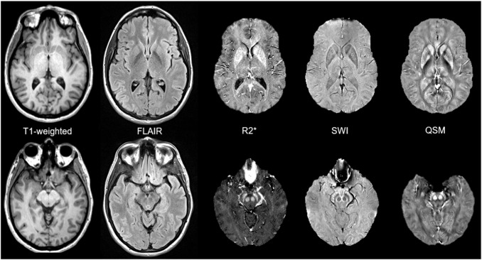
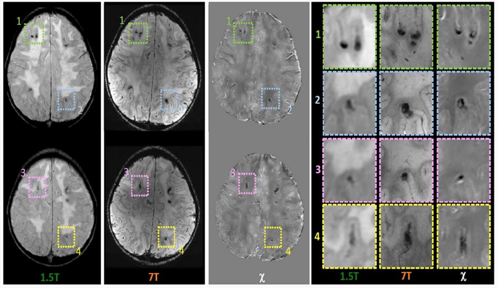
ABSTRACT: Background: The brainstem contains grey matter nuclei and white matter tracts to be identified in clinical practice. The small size and the low contrast among them make their in vivo visualisation challenging using conventional magnetic resonance imaging (MRI) sequences at high magnetic field strengths. Combining higher spatial resolution, signal- and contrast-to-noise ratio and sensitivity to magnetic susceptibility (χ), susceptibility-weighted 7-T imaging could improve the assessment of brainstem anatomy.
Methods: We acquired high-resolution 7-T MRI of the brainstem in a 46-year-old female healthy volunteer (using a three-dimensional multi-echo gradient-recalled-echo sequence; spatial resolution 0.3 × 0.3 × 1.2 mm3) and in a brainstem sample from a 48-year-old female body donor that was sectioned and stained. Images were visually assessed; nuclei and tracts were labelled and named according to the official nomenclature.
Results: This in vivo imaging revealed structures usually evaluated through light microscopy, such as the accessory olivary nuclei, oculomotor nucleus and the medial longitudinal fasciculus. Some fibre tracts, such as the medial lemniscus, were visible for most of their course. Overall, in in vivo acquisitions, χ and frequency maps performed better than T2*-weighted imaging and allowed for the evaluation of a greater number of anatomical structures. All the structures identified in vivo were confirmed by the ex vivo imaging and histology.
Conclusions: The use of multi-echo GRE sequences at 7 T allowed the visualisation of brainstem structures that are not visible in detail at conventional magnetic field and opens new perspectives in the diagnostic and therapeutical approach to brain disorders.
Relevance statement: In vivo MR imaging at UHF provides detailed anatomy of CNS substructures comparable to that obtained with histology. Anatomical details are fundamentals for diagnostic purposes but also to plan a direct targeting for a minimally invasive brain stimulation or ablation.
Cencini M, Lancione M, Pasquariello R, Peretti L, Pirkl CM, Schulte RF, Buonincontri G, Arduino A, Zilberti L, Biagi L, Tosetti M
Fast high-resolution electric properties tomography using three-dimensional quantitative transient-state imaging-based water fraction estimation
NMR in Biomedicine, e5039, 2023
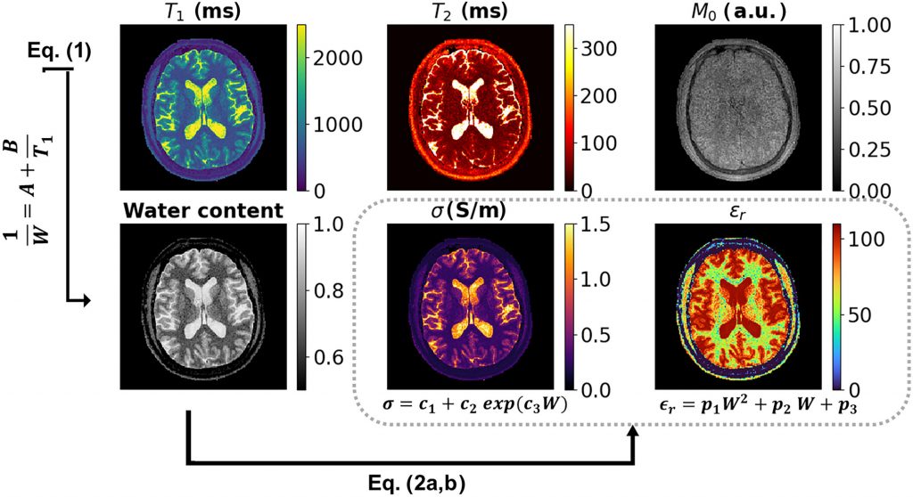
ABSTRACT: In this study, we aimed to develop a fast and robust high-resolution technique for clinically feasible electrical properties tomography based on water content maps (wEPT) using Quantitative Transient-state Imaging (QTI), a multiparametric transient state-based method that is similar to MR fingerprinting. Compared with the original wEPT implementation based on standard spin-echo acquisition, QTI provides robust electrical properties quantification towards B1+ inhomogeneities and full quantitative relaxometry data. To validate the proposed approach, 3D QTI data of 12 healthy volunteers were acquired on a 1.5 T scanner. QTI-provided T1 maps were used to compute water content maps of the tissues using an empirical relationship based on literature ex-vivo measurements. Assuming that electrical properties are modulated mainly by tissue water content, the water content maps were used to derive electrical conductivity and relative permittivity maps. The proposed technique was compared with a conventional phase-only Helmholtz EPT (HH-EPT) acquisition both within whole white matter, gray matter, and cerebrospinal fluid masks, and within different white and gray matter subregions. In addition, QTI-based wEPT was retrospectively applied to four multiple sclerosis adolescent and adult patients, compared with conventional contrast-weighted imaging in terms of lesion delineation, and quantitatively assessed by measuring the variation of electrical properties in lesions. Results obtained with the proposed approach agreed well with theoretical predictions and previous in vivo findings in both white and gray matter. The reconstructed maps showed greater anatomical detail and lower variability compared with standard phase-only HH-EPT. The technique can potentially improve delineation of pathology when compared with conventional contrast-weighted imaging and was able to detect significant variations in lesions with respect to normal-appearing tissues. In conclusion, QTI can reliably measure conductivity and relative permittivity of brain tissues within a short scan time, opening the way to the study of electric properties in clinical settings.
Donatelli G, Cecchi P, Migaleddu G, Cencini M, Frumento P, D’Amelio C, Peretti L, Buonincontri G, Pasquali L, Tosetti M, Cosottini M, Costagli M
Quantitative T1 mapping detects blood–brain barrier breakdown in apparently non-enhancing multiple sclerosis lesions
NeuroImage: Clinical, 40:103509, 2023

ABSTRACT: Objectives: The disruption of the blood–brain barrier (BBB) is a key and early feature in the pathogenesis of demyelinating multiple sclerosis (MS) lesions and has been neuropathologically demonstrated in both active and chronic plaques. The local overt BBB disruption in acute demyelinating lesions is captured as signal hyperintensity in post-contrast T1-weighted images because of the contrast-related shortening of the T1 relaxation time. On the contrary, the subtle BBB disruption in chronic lesions is not visible at conventional radiological evaluation but it might be of clinical relevance. Indeed, persistent, subtle BBB leakage might be linked to low-grade inflammation and plaque evolution. Here we hypothesised that 3D Quantitative Transient-state Imaging (QTI) was able to reveal and measure T1 shortening (ΔT1) reflecting small amounts of contrast media leakage in apparently non-enhancing lesions (ANELs). Materials and methods: Thirty-four patients with relapsing remitting MS were included in the study. All patients underwent a 3 T MRI exam of the brain including conventional sequences and QTI acquisitions (1.1 mm isotropic voxel) performed both before and after contrast media administration. For each patient, a ΔT1 map was obtained via voxel-wise subtraction of pre- and post- contrast QTI-derived T1 maps. ΔT1 values measured in ANELs were compared with those recorded in enhancing lesions and in the normal appearing white matter. A reference distribution of ΔT1 in the white matter was obtained from datasets acquired in 10 non-MS patients with unrevealing MR imaging. Results: Mean ΔT1 in ANELs (57.45 ± 48.27 ms) was significantly lower than in enhancing lesions (297.71 ± 177.52 ms; p < 0. 0001) and higher than in the normal appearing white matter (36.57 ± 10.53 ms; p < 0.005). Fifty-two percent of ANELs exhibited ΔT1 higher than those observed in the white matter of non-MS patients. Conclusions: QTI-derived quantitative ΔT1 mapping enabled to measure contrast-related T1 shortening in ANELs. ANELs exhibiting ΔT1 values that deviate from the reference distribution in non-MS patients may indicate persistent, subtle, BBB disruption. Access to this information may be proved useful to better characterise pathology and objectively monitor disease activity and response to therapy.
Peretti L, Donatelli G, Cencini M, Cecchi P, Buonincontri G, Cosottini M, Tosetti M, Costagli M
Generating Synthetic Radiological Images with PySynthMRI: An Open-Source Cross-Platform Tool
Tomography, 9(5):1723-1733, 2023
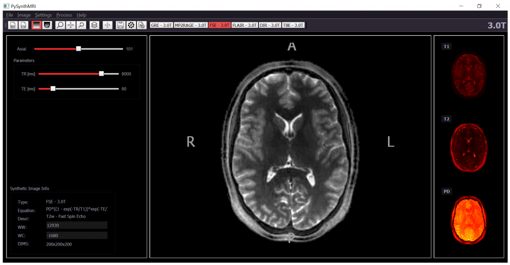
ABSTRACT: Synthetic MR Imaging allows for the reconstruction of different image contrasts from a single acquisition, reducing scan times. Commercial products that implement synthetic MRI are used in research. They rely on vendor-specific acquisitions and do not include the possibility of using custom multiparametric imaging techniques. We introduce PySynthMRI, an open-source tool with a user-friendly interface that uses a set of input images to generate synthetic images with diverse radiological contrasts by varying representative parameters of the desired target sequence, including the echo time, repetition time and inversion time(s). PySynthMRI is written in Python 3.6, and it can be executed under Linux, Windows, or MacOS as a python script or an executable. The tool is free and open source and is developed while taking into consideration the possibility of software customization by the end user. PySynthMRI generates synthetic images by calculating the pixelwise signal intensity as a function of a set of input images (e.g., T1 and T2 maps) and simulated scanner parameters chosen by the user via a graphical interface. The distribution provides a set of default synthetic contrasts, including T1w gradient echo, T2w spin echo, FLAIR and Double Inversion Recovery. The synthetic images can be exported in DICOM or NiFTI format. PySynthMRI allows for the fast synthetization of differently weighted MR images based on quantitative maps. Specialists can use the provided signal models to retrospectively generate contrasts and add custom ones. The modular architecture of the tool can be exploited to add new features without impacting the codebase.
Bosco P, Lancione M, Retico A, Nigri A, Aquino D, Baglio F, Carne I, Ferraro S, Giulietti G, Napolitano A, Palesi F, Pavone L, Savini G, Tagliavini F, Bruzzone MG, Gandini Wheeler-Kingshott CAM, Tosetti M, Biagi L, the RIN-Neuroimaging Network
Quality assessment, variability and reproducibility of anatomical measurements derived from T1-weighted brain imaging: The RIN–Neuroimaging Network case study
Physica Medica, 110:102577, 2023
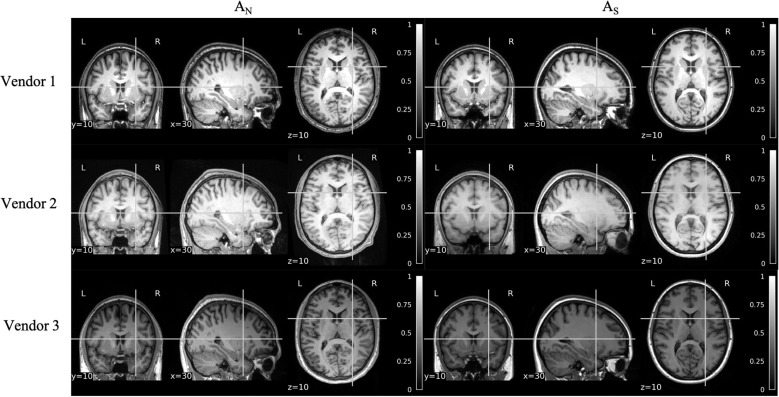
ABSTRACT: Initiatives for the collection of harmonized MRI datasets are growing continuously, opening questions on the reliability of results obtained in multi-site contexts.
Here we present the assessment of the brain anatomical variability of MRI-derived measurements obtained from T1-weighted images, acquired according to the Standard Operating Procedures, promoted by the RIN-Neuroimaging Network. A multicentric dataset composed of 77 brain T1w acquisitions of young healthy volunteers (mean age = 29.7 ± 5.0 years), collected in 15 sites with MRI scanners of three different vendors, was considered. Parallelly, a dataset of 7 “traveling” subjects, each undergoing three acquisitions with scanners from different vendors, was also used. Intra-site, intra-vendor, and inter-site variabilities were evaluated in terms of the percentage standard deviation of volumetric and cortical thickness measures. Image quality metrics such as contrast-to-noise and signal-to-noise ratio in gray and white matter were also assessed for all sites and vendors.
The results showed a measured global variability that ranges from 11% to 19% for subcortical volumes and from 3% to 10% for cortical thicknesses. Univariate distributions of the normalized volumes of subcortical regions, as well as the distributions of the thickness of cortical parcels appeared to be significantly different among sites in 8 subcortical (out of 17) and 21 cortical (out of 68) regions of i nterest in the multicentric study.
The Bland-Altman analysis on “traveling” brain measurements did not detect systematic scanner biases even though a multivariate classification approach was able to classify the scanner vendor from brain measures with an accuracy of 0.60 ± 0.14 (chance level 0.33).
Cuña EG, Schulz H, Tuzzi E, Biagi L, Bosco P, García-Fontes M, Mattos J, Tosetti M, Engelmann J, Scheffler K, Hagberg GE
Simulated and experimental phantom data for multi-center quality assurance of quantitative susceptibility maps at 3 T, 7 T and 9.4 T
Physica Medica, 110:102590, 2023
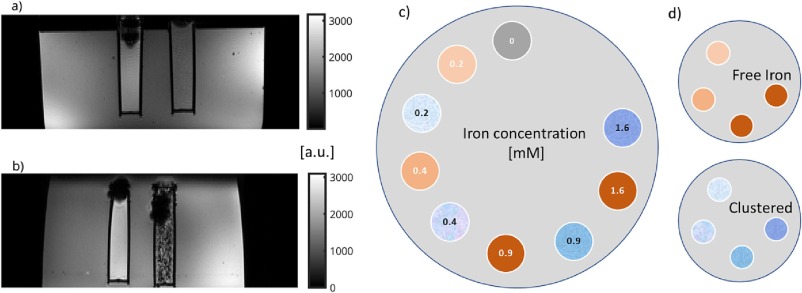
ABSTRACT: Purpose: To develop methods for quality assurance of quantitative susceptibility mapping (QSM) using MRI at different magnetic field strengths, and scanners, using different MR-sequence protocols, and post-processing pipelines. Methods: We built a custom phantom based on iron in two forms: homogeneous susceptibility (‘free iron’) and with fine-scaled variations in susceptibility (‘clustered iron’) at different iron concentrations. The phantom was measured at 3.0 T (two scanners), 7.0 T and 9.4 T using multi-echo, gradient echo acquisition sequences. A digital phantom analogue to the iron-phantom, tailored to obtain similar results as in experimentation was developed, with similar geometry and susceptibility values. Morphology enabled dipole inversion was applied to the phase images to obtain QSM for experimental and simulated data using the MEDI + 0 approach for background regularization. Results: Across all scanners, QSM-values showed a linear increase with iron concentrations. The QSM-relaxivity was 0.231 ± 0.047 ppm/mM for free and 0.054 ± 0.013 ppm/mM for clustered iron, with adjusted determination coefficients (DoC) ≥ 0.87. Similarly, the simulations yielded linear increases (DoC ≥ 0.99). In both the experimental and digital phantoms, the estimated molar susceptibility was lower with clustered iron, because clustering led to highly localized field effects. Conclusion: Our iron phantom can be used to evaluate the capability of QSM to detect local variations in susceptibility across different field strengths, when using different MR-sequence protocols. The devised simulation method captures the effect of iron clustering in QSM as seen experimentally and could be used in the future to optimize QSM processing pipelines and achieve higher accuracy for local field effects, as also seen in Alzheimer’s beta-amyloid plaques.
Biagi L, Tosetti M, Crespi SA, and Morrone MC
Development of BOLD response to motion in human infants
Journal of Neuroscience, JN-RM-0837-22, 2023
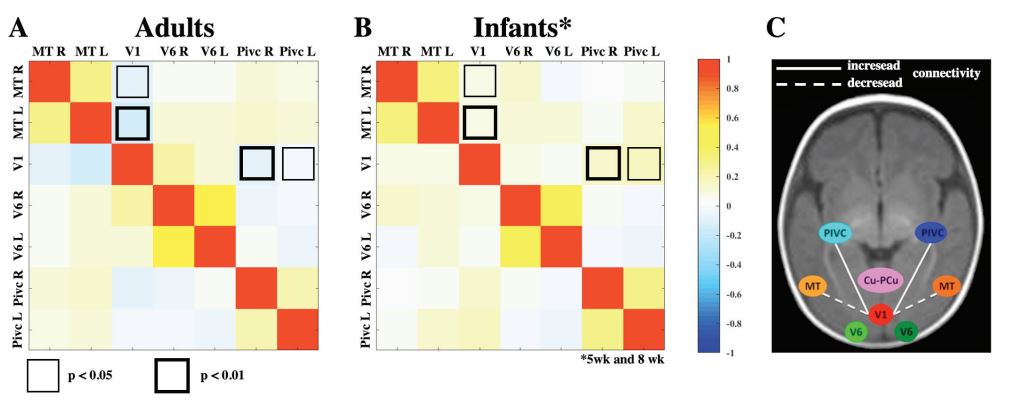
ABSTRACT: Behavioral studies suggest that motion perception is rudimentary at birth and matures steadily over the first few years. We demonstrated previously that the major cortical associative areas serving motion processing, like MT+, V6 and PVICS in adults, show selective responses to coherent flow in 8-week-old infants. Here we study the BOLD response to the same motion stimuli in 5-week-old infants (four females and four males) and compare the maturation between these two ages. The results show that MT+ and PVIC areas show a similar motion response at 5 and 8 weeks, while response in the V6 area shows a reduced BOLD response to motion at 5 weeks and Cuneus associative areas are not identifiable at this young age. In infants and in adults, V1 does not show a selectivity for coherent motion, but shows a very fast development between 5 and 8 weeks of age in response to appearance of motion stimuli. Resting-state correlations demonstrate adult-like functional connectivity between the motion-selective associative areas, but not between primary cortex and temporo-occipital and posterior-insular cortices. The results are consistent with a differential developmental trajectory of motion area respect to other occipital regions, probably reflecting also a different development trajectory of the central and peripheral visual field.
Bombonato C, Cipriano E, Pecini C, Casalini C, Bosco P, Podda I, Tosetti M, Biagi L, Chilosi AM
Relationship among Connectivity of the Frontal Aslant Tract, Executive Functions, and Speech and Language Impairment in Children with Childhood Apraxia of Speech
Brain Sciences, 13:78, 2023
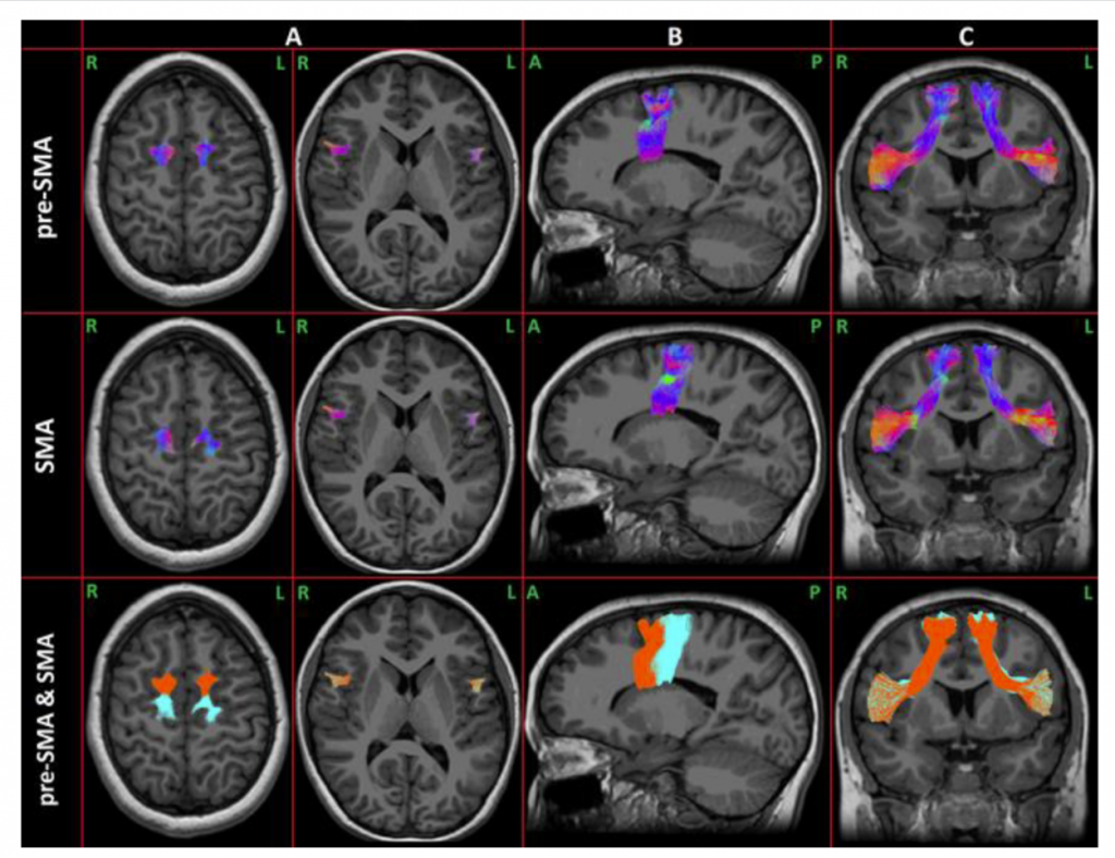
ABSTRACT: Childhood apraxia of speech (CAS) is a subtype of motor speech disorder usually co-occurring with language impairment. A supramodal processing difficulty, involving executive functions (EFs), might contribute to the cognitive endophenotypes and behavioral manifestations. The present study aimed to profile the EFs in CAS, investigating the relationship between EFs, speech and language severity, and the connectivity of the frontal aslant tract (FAT), a white matter tract involved in both speech and EFs. A total of 30 preschool children with CAS underwent speech, language, and EF assessments and brain MRIs. Their FAT connectivity metrics were compared to those of 30 children without other neurodevelopmental disorders (NoNDs), who also underwent brain MRIs. Alterations in some basic EF components were found. Inhibition and working memory correlated with speech and language severity. Compared to NoND children, a weak, significant reduction in fractional anisotropy (FA) in the left presupplementary motor area (preSMA) FAT component was found. Only speech severity correlated and predicted FA values along with the FAT in both of its components, and visual-spatial working memory moderated the relationship between speech severity and FA in the left SMA. Our study supports the conceptualization of a composite and complex picture of CAS, not limited to the speech core deficit, but also involving high-order cognitive skills.
2022
Palesi F, Nigri A, Gianeri R, Aquino D, Redolfi A, Biagi L, Carne I, De Francesco S, Ferraro S, Martucci P, Medina JP, Napolitano A, Pirastru A, Baglio F, Tagliavini F, Bruzzone MG, Tosetti M, Gandini Wheeler-Kingshott CAM, the RIN-Neuroimaging Network
MRI data quality assessment for the RIN – Neuroimaging Network using the ACR phantoms
Physica Medica, 104:93-100, 2022

ABSTRACT: Purpose: Generating big-data is becoming imperative with the advent of machine learning. RIN-Neuroimaging Network addresses this need by developing harmonized protocols for multisite studies to identify quantitative MRI (qMRI) biomarkers for neurological diseases. In this context, image quality control (QC) is essential. Here, we present methods and results of how the RIN performs intra- and inter-site reproducibility of geometrical and image contrast parameters, demonstrating the relevance of such QC practice.
Methods: American College of Radiology (ACR) large and small phantoms were selected. Eighteen sites were equipped with a 3T scanner that differed by vendor, hardware/software versions, and receiver coils. The standard ACR protocol was optimized (in-plane voxel, post-processing filters, receiver bandwidth) and repeated monthly. Uniformity, ghosting, geometric accuracy, ellipse’s ratio, slice thickness, and high-contrast detectability tests were performed using an automatic QC script.
Results: Measures were mostly within the ACR tolerance ranges for both T1- and T2-weighted acquisitions, for all scanners, regardless of vendor, coil, and signal transmission chain type. All measurements showed good reproducibility over time. Uniformity and slice thickness failed at some sites. Scanners that upgraded the signal transmission chain showed a decrease in geometric distortion along the slice encoding direction. Inter-vendor differences were observed in uniformity and geometric measurements along the slice encoding direction (i.e. ellipse’s ratio).
Conclusions: Use of the ACR phantoms highlighted issues that triggered interventions to correct performance at some sites and to improve the longitudinal stability of the scanners. This is relevant for establishing precision levels for future multisite studies of qMRI biomarkers.
Lancione M, Bosco P, Costagli M, Nigri A, Aquino D, Carne I, Ferraro S, Giulietti G, Napolitano A, Palesi F, Pavone L, Pirastru A, Savini G, Tagliavini F, Bruzzone MG, Gandini Wheeler-Kingshott CAM, Tosetti M, Biagi L, the RIN-Neuroimaging Network
Multi-centre and multi-vendor reproducibility of a standardized protocol for quantitative susceptibility Mapping of the human brain at 3T
Physica Medica, 103:37-45, 2022

ABSTRACT: Quantitative Susceptibility Mapping (QSM) is an MRI-based technique allowing the non-invasive quantification of iron content and myelination in the brain. The RIN – Neuroimaging Network established an optimized and harmonized protocol for QSM across ten sites with 3T MRI systems from three different vendors to enable multicentric studies. The assessment of the reproducibility of this protocol is crucial to establish susceptibility as a quantitative biomarker. In this work, we evaluated cross-vendor reproducibility in a group of six traveling brains. Then, we recruited fifty-one volunteers and measured the variability of QSM values in a cohort of healthy subjects scanned at different sites, simulating a multicentric study. Both voxelwise and Region of Interest (ROI)-based analysis on cortical and subcortical gray matter were performed.
The traveling brain study yielded high structural similarity (~0.8) and excellent reproducibility comparing maps acquired on scanners from two different vendors. Depending on the ROI, we reported a quantification error
ranging from 0.001 to 0.017 ppm for the traveling brains. In the cohort of fifty-one healthy subjects scanned at
nine different sites, the ROI-dependent variability of susceptibility values, of the order of 0.005–0.025 ppm, was
comparable to the result of the traveling brain experiment.
The harmonized QSM protocol of the RIN – Neuroimaging Network provides a reliable quantification of
susceptibility in both cortical and subcortical gray matter regions and it is ready for multicentric and longitudinal
clinical studies in neurological and pychiatric diseases.
Cervelli R, Cencini M, Cacciato Insilla A, Aringhieri G, Boggi U, Campani D, Tosetti M, Crocetti L
Ex-vivo human pancreatic specimen evaluation by 7 Tesla MRI: a prospective radiological-pathological correlation study
Radiol Med, 127:950–959, 2022

ABSTRACT: To compare the characteristics detected by 7Tesla (7 T) MR and the histological composition of ex-vivo specimens from lesions diagnosed at preoperative CT scan as Pancreatic Ductal Adenocarcinoma (PDAC).
Ten pancreatic specimens were examined. The 7 T imaging protocol included both morphologic and quantitative sequences; the latter was acquired by conventional methods and a novel multiparametric method, the magnetic resonance fingerprinting (MRF) sequence. Two radiologists reviewed the images to: (1) evaluate the quality of the morphological and quantitative sequences by assigning an “image consistency score” on a 4-point scale; (2) identify the lesion, recording its characteristics; (3) perform the quantitative analysis on “target lesion” and “non target tissue”. Finally, the specimen was analysed by two pathologists.
Seven out of 10 lesions were PDAC, 2/10 were biliary carcinomas, whereas one lesion was an ampullary adenocarcinoma. The quality of the morphological sequences was judged “excellent”. The “image consistency score” for the conventional quantitative sequences and MRF were 2.8 ± 0.42 and 2.9 ± 0.57; the “overall MR examination score” was 3.5 ± 0.53. A statistical correlation was found between the relaxation time values of conventional and MRF T1-weighted sequences (p < 0.0001), as well as between conventional and MRF fat- and water-fraction maps (p < 0.05). The “target lesion” and “non target tissue” relaxation time values were statistically different according to conventional T1-, T2-weighted, and MRF T1-weighted sequences.
Conventional T1-, T2-weighted sequences and MRF derived relaxometries may be useful in differentiating between tumour and non-target pancreatic tissue. Moreover, the MRF sequence can be used to obtain reliable relaxation time data.
Lancione M, Donatelli G, Del Prete E, Campese N, Frosini D, Cencini M, Costagli M, Biagi L, Lucchi G, Tosetti M, Godani M, Arnaldi D, Terzaghi M, Provini F, Pacchetti C, Cortelli P, Bonanni E, Ceravolo R, Cosottini M
Evaluation of iron overload in Nigrosome 1 via Quantitative Susceptibility Mapping as a progression biomarker in prodromal stages of synucleinopathies
NeuroImage, 260:119454, 2022

ABSTRACT: Idiopathic rapid eye movement (REM) sleep behavior disorder (iRBD) is a prodromal stage of α-synucleinopathies, such as Parkinson’s disease (PD), which are characterized by the loss of dopaminergic neurons in substantia nigra, associated with abnormal iron load. The assessment of presymptomatic biomarkers predicting the onset of neurodegenerative disorders is critical for monitoring early signs, screening patients for neuroprotective clinical trials and understanding the causal relationship between iron accumulation processes and disease development. Here, we used Quantitative Susceptibility Mapping (QSM) and 7T MRI to quantify iron deposition in Nigrosome 1 (N1) in early PD (ePD) patients, iRBD patients and healthy controls and investigated group differences and correlation with disease progression. We evaluated the radiological appearance of N1 and analyzed its iron content in 35 ePD, 30 iRBD patients and 14 healthy controls via T2*-weighted sequences and susceptibility (χ) maps. N1 regions of interest (ROIs) were manually drawn on control subjects and warped onto a study-specific template to obtain probabilistic N1 ROIs. For each subject the N1 with the highest mean χ was considered for statistical analysis. The appearance of N1 was rated pathological in 45% of iRBD patients. ePD patients showed increased N1 χ compared to iRBD patients and HC but no correlation with disease duration, indicating that iron load remains stable during the early stages of disease progression. Although no difference was reported in iron content between iRBD and HC, N1 χ in the iRBD group increases as the disease evolves. QSM can reveal temporal changes in N1 iron content and its quantification may represent a valuable presymptomatic biomarker to assess neurodegeneration in the prodromal stages of PD.
Nigri A, Ferraro S, Gandini Wheeler-Kingshott CAM, Tosetti M, Redolfi A, Forloni G, D’Angelo E, Aquino D, Biagi L, Bosco P, et al., the RIN-Neuroimaging Network
Quantitative MRI Harmonization to Maximize Clinical Impact: The RIN-Neuroimaging Network
Frontiers in Neurology, 13:855125, 2022

ABSTRACT: Neuroimaging studies often lack reproducibility, one of the cardinal features of the scientific method. Multisite collaboration initiatives increase sample size and limit methodological flexibility, therefore providing the foundation for increased statistical power and generalizable results. However, multisite collaborative initiatives are inherently limited by hardware, software, and pulse and sequence design heterogeneities of both clinical and preclinical MRI scanners and the lack of benchmark for acquisition protocols, data analysis, and data sharing. We present the overarching vision that yielded to the constitution of RIN-Neuroimaging Network, a national consortium dedicated to identifying disease and subject-specific in-vivo neuroimaging biomarkers of diverse neurological and neuropsychiatric conditions. This ambitious goal needs efforts toward increasing the diagnostic and prognostic power of advanced MRI data. To this aim, 23 Italian Scientific Institutes of Hospitalization and Care (IRCCS), with technological and clinical specialization in the neurological and neuroimaging field, have gathered together. Each IRCCS is equipped with high- or ultra-high field MRI scanners (i.e., ≥3T) for clinical or preclinical research or has established expertise in MRI data analysis and infrastructure. The actions of this Network were defined across several work packages (WP). A clinical work package (WP1) defined the guidelines for a minimum standard clinical qualitative MRI assessment for the main neurological diseases. Two neuroimaging technical work packages (WP2 and WP3, for clinical and preclinical scanners) established Standard Operative Procedures for quality controls on phantoms as well as advanced harmonized quantitative MRI protocols for studying the brain of healthy human participants and wild type mice. Under FAIR principles, a web-based e-infrastructure to store and share data across sites was also implemented (WP4). Finally, the RIN translated all these efforts into a large-scale multimodal data collection in patients and animal models with dementia (i.e., case study). The RIN-Neuroimaging Network can maximize the impact of public investments in research and clinical practice acquiring data across institutes and pathologies with high-quality and highly-consistent acquisition protocols, optimizing the analysis pipeline and data sharing procedures.
Kurzawski JW, Lunghi C, Biagi L, Tosetti M, Morrone MC, Binda P
Short-term plasticity in the human visual thalamus
Elife, 11:74565, 2022

ABSTRACT: While there is evidence that the visual cortex retains a potential for plasticity in adulthood, less is known about the subcortical stages of visual processing. Here we asked whether short-term ocular dominance plasticity affects the human visual thalamus. We addressed this question in normally sighted adult humans, using ultra-high field (7T) magnetic resonance imaging combined with the paradigm of short-term monocular deprivation. With this approach, we previously demonstrated transient shifts of perceptual eye dominance and ocular dominance in visual cortex (Binda et al., 2018). Here we report evidence for short-term plasticity in the ventral division of the pulvinar (vPulv), where the deprived eye representation was enhanced over the non-deprived eye. This ventral-pulvinar plasticity was similar as previously seen in visual cortex and it was correlated with the ocular dominance shift measured behaviorally. In contrast, there was no effect of monocular deprivation in two adjacent thalamic regions: dorsal pulvinar (dPulv), and Lateral Geniculate Nucleus (LGN). We conclude that the visual thalamus retains potential for short-term plasticity in adulthood; the plasticity effect differs across thalamic subregions, possibly reflecting differences in their cortico-fugal connectivity.
Pirkl CM, Cencini M, Kurzawski JW, Waldmannstetter D, Li H, Sekuboyina A, Endt S, Peretti L, Donatelli G, Pasquariello R, Costagli M, Buonincontri G, Tosetti M, Menzel MI, Menze BH
Learning residual motion correction for fast and robust 3D multiparametric MRI
Medical Image Analysis, 77:102387, 2022

ABSTRACT: Voluntary and involuntary patient motion is a major problem for data quality in clinical routine of Magnetic Resonance Imaging (MRI). It has been thoroughly investigated and, yet it still remains unresolved. In quantitative MRI, motion artifacts impair the entire temporal evolution of the magnetization and cause errors in parameter estimation. Here, we present a novel strategy based on residual learning for retrospective motion correction in fast 3D whole-brain multiparametric MRI. We propose a 3D multiscale convolutional neural network (CNN) that learns the non-linear relationship between the motion-affected quantitative parameter maps and the residual error to their motion-free reference. For supervised model training, despite limited data availability, we propose a physics-informed simulation to generate self-contained paired datasets from a priori motion-free data. We evaluate motion-correction performance of the proposed method for the example of 3D Quantitative Transient-state Imaging at 1.5T and 3T. We show the robustness of the motion correction for various motion regimes and demonstrate the generalization capabilities of the residual CNN in terms of real-motion in vivo data of healthy volunteers and clinical patient cases, including pediatric and adult patients with large brain lesions. Our study demonstrates that the proposed motion correction outperforms current state of the art, reliably providing a high, clinically relevant image quality for mild to pronounced patient movements. This has important implications in clinical setups where large amounts of motion affected data must be discarded as they are rendered diagnostically unusable.
Fujita S, Cencini M, Buonincontri G, Takei N, Schulte RF, Fukunaga I, Uchida W, Hagiwara A, Kamagata K, Hagiwara Y, Matsuyama Y, Abe O, Tosetti M, Aoki S
Simultaneous relaxometry and morphometry of human brain structures with 3D magnetic resonance fingerprinting: a multicenter, multiplatform, multifield-strength study
Cerebral Cortex, 33:729-739, 2022
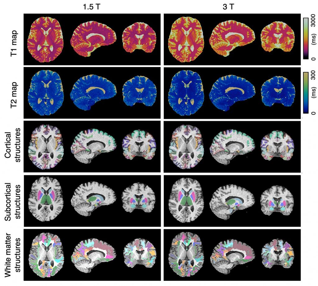
ABSTRACT: Relaxation times and morphological information are fundamental magnetic resonance imaging-derived metrics of the human brain that reflect the status of the underlying tissue. Magnetic resonance fingerprinting (MRF) enables simultaneous acquisition of T1 and T2 maps inherently aligned to the anatomy, allowing whole-brain relaxometry and morphometry in a single scan. In this study, we revealed the feasibility of 3D MRF for simultaneous brain structure-wise morphometry and relaxometry. Comprehensive test–retest scan analyses using five 1.5-T and three 3.0-T systems from a single vendor including different scanner types across 3 institutions demonstrated that 3D MRF-derived morphological information and relaxation times are highly repeatable at both 1.5 T and 3.0 T. Regional cortical thickness and subcortical volume values showed high agreement and low bias across different field strengths. The ability to acquire a set of regional T1, T2, thickness, and volume measurements of neuroanatomical structures with high repeatability and reproducibility facilitates the ability of longitudinal multicenter imaging studies to quantitatively monitor changes associated with underlying pathologies, disease progression, and treatments.
Lancione M, Cencini M, Costagli M, Donatelli G, Tosetti M, Giannini G, Zangaglia R, Calandra-Buonaura G, Pacchetti C, Cortelli P, Cosottini M
Diagnostic accuracy of Quantitative Susceptibility Mapping in Multiple System Atrophy: the impact of echo time and the potential of histogram analysis
NeuroImage: Clinical, 34:102989, 2022

ABSTRACT: The non-invasive quantification of iron stores via Quantitative Susceptibility Mapping (QSM) could play an important role in the diagnosis and the differential diagnosis of atypical Parkinsonisms. However, the susceptibility (χ) values measured via QSM depend on echo time (TE). This effect relates to the microstructural organization within the voxel, whose composition can be altered by the disease. Moreover, pathological iron deposition in a brain area may not be spatially uniform, and conventional Region of Interest (ROI)-based analysis may fail in detecting alterations. Therefore, in this work we evaluated the impact of echo time on the diagnostic accuracy of QSM on a population of patients with Multiple System Atrophy (MSA) of either Parkinsonian (MSAp) or cerebellar (MSAc) phenotypes. In addition, we tested the potential of histogram analysis to improve QSM classification accuracy.
We enrolled 32 patients (19 MSAp and 13 MSAc) and 16 healthy controls, who underwent a 7T MRI session including a gradient-recalled multi-echo sequence for χ mapping. Nine histogram features were extracted from the χ maps computed for each TE in atlas-based ROIs covering deep brain nuclei, and compared among groups.
Alterations of susceptibility distribution were found in the Putamen, Substantia Nigra, Globus Pallidus and Caudate Nucleus for MSAp and in the Substantia Nigra and Dentate Nucleus for MSAc. Increased iron deposition was observed in a larger number of ROIs for the two shortest TEs and the standard deviation, the 75th and the 90th percentile were the most informative features yielding excellent diagnostic accuracy with area under the ROC curve > 0.9.
In conclusion, short TEs may enhance QSM diagnostic performances, as they can capture variations in rapidly-decaying contributions of high χ sources. The analysis of histogram features allowed to reveal fine heterogeneities in the spatial distribution of susceptibility alteration, otherwise undetected by a simple evaluation of ROI χ mean values.
2021
Lancione M, Costagli M, Handjaras G, Tosetti M, Ricciardi E, Pietrini P, Cecchetti L
Complementing canonical fMRI with functional Quantitative Susceptibility Mapping (fQSM) in modern neuroimaging research
NeuroImage, 244:118574, 2021

ABSTRACT: Functional Quantitative Susceptibility Mapping (fQSM) allows for the quantitative measurement of time-varying magnetic susceptibility across cortical and subcortical brain structures with a potentially higher spatial specificity than conventional fMRI. While the usefulness of fQSM with General Linear Model and “On/Off” paradigms has been assessed, little is known about the potential applications and limitations of this technique in more sophisticated experimental paradigms and analyses, such as those currently used in modern neuroimaging.
To thoroughly characterize fQSM activations, here we used 7T MRI, tonotopic mapping, as well as univariate (i.e., GLM and population Receptive Field) and multivariate (Representational Similarity Analysis; RSA) analyses.
Although fQSM detected less tone-responsive voxels than fMRI, they were more consistently localized in gray matter. Also, the majority of active gray matter voxels exhibited negative fQSM response, signaling the expected oxyhemoglobin increase, whereas positive fQSM activations were mainly in white matter. Though fMRI- and fQSM-based tonotopic maps were overall comparable, the representation of frequency tunings in tone-sensitive regions was significantly more balanced for fQSM. Lastly, RSA revealed that frequency information from the auditory cortex could be successfully retrieved by using either methods.
Overall, fQSM produces complementary results to conventional fMRI, as it captures small-scale variations in the activation pattern which inform multivariate measures. Although positive fQSM responses deserve further investigation, they do not impair the interpretation of contrasts of interest. The quantitative nature of fQSM, its spatial specificity and the possibility to simultaneously acquire canonical fMRI support the use of this technique for longitudinal and multicentric studies and pre-surgical mapping.
Düzel E, Costagli M, Donatelli G, Speck O, Cosottini M
Studying Alzheimer disease, Parkinson disease, and amyotrophic lateral sclerosis with 7-T magnetic resonance
European radiology experimental, 5(1):1-17, 2021

ABSTRACT: Ultra-high-field (UHF) magnetic resonance (MR) scanners, that is, equipment operating at static magnetic field of 7 tesla (7 T) and above, enable the acquisition of data with greatly improved signal-to-noise ratio with respect to conventional MR systems (e.g., scanners operating at 1.5 T and 3 T). The change in tissue relaxation times at UHF offers the opportunity to improve tissue contrast and depict features that were previously inaccessible. These potential advantages come, however, at a cost: in the majority of UHF-MR clinical protocols, potential drawbacks may include signal inhomogeneity, geometrical distortions, artifacts introduced by patient respiration, cardiac cycle, and motion. This article reviews the 7 T MR literature reporting the recent studies on the most widespread neurodegenerative diseases: Alzheimer’s disease, Parkinson’s disease, and amyotrophic lateral sclerosis.
Ambrosi P, Costagli M, Kuruoğlu EE, Biagi L, Buonincontri G, Tosetti M
Modeling brain connectivity dynamics in functional magnetic resonance imaging via particle filtering
Brain Informatics, 8:19, 2021

ABSTRACT: Interest in the studying of functional connections in the brain has grown considerably in the last decades, as many studies have pointed out that alterations in the interaction among brain areas can play a role as markers of neurological diseases. Most studies in this field treat the brain network as a system of connections stationary in time, but dynamic features of brain connectivity can provide useful information, both on physiology and pathological conditions of the brain. In this paper, we propose the application of a computational methodology, named Particle Filter (PF), to study non-stationarities in brain connectivity in functional Magnetic Resonance Imaging (fMRI). The PF algorithm estimates time-varying hidden parameters of a first-order linear time-varying Vector Autoregressive model (VAR) through a Sequential Monte Carlo strategy. On simulated time series, the PF approach effectively detected and enabled to follow time-varying hidden parameters and it captured causal relationships among signals. The method was also applied to real fMRI data, acquired in presence of periodic tactile or visual stimulations, in different sessions. On these data, the PF estimates were consistent with current knowledge on brain functioning. Most importantly, the approach enabled to detect statistically significant modulations in the cause-effect relationship between brain areas, which correlated with the underlying visual stimulation pattern presented during the acquisition.
Benedetto A, Binda P, Costagli M, Tosetti M, Morrone MC
Predictive visuo-motor communication through neural oscillations
Current Biology, 31(15):3401-3408.e4, 2021

ABSTRACT: The mechanisms coordinating action and perception over time are poorly understood. The sensory cortex needs to prepare for upcoming changes contingent on action, and this requires temporally precise communication that takes into account the variable delays between sensory and motor processing. Several theorists have proposed synchronization of the endogenous oscillatory activity observed in most regions of the brain as the basis for an efficient and flexible communication protocol between distal brain areas a concept known as “communication through coherence”. Synchronization of endogenous oscillations occurs after a salient sensory stimulus, such as a flash or a sound, and after a voluntary action, and this directly impacts perception, causing performance to oscillate rhythmically over time. Here we introduce a novel fMRI paradigm to probe the neural sources of oscillations, based on the concept of perturbative signals, which overcomes the low temporal resolution of BOLD signals. The assumption is that a synchronized endogenous rhythm will modulate cortical excitability rhythmically, which should be reflected in the BOLD responses to brief stimuli presented at different phases of the oscillation cycle. We record rhythmic oscillations of V1 BOLD synchronized by a simple voluntary action, in phase with behaviorally measured oscillations in visual sensitivity in the theta range. The functional connectivity between V1 and M1 also oscillates at the same rhythm. By demonstrating oscillatory temporal coupling between primary motor and sensory cortices, our results strongly implicate communication through coherence to achieve precise coordination and to encode sensory-motor timing.
Leombruni O, Annovi A, Giannetti P, Biesuz NV, Roda C, Calvetti M, Piendibene M, Peretti L, Cencini M, Tosetti M, Buonincontri G
Pattern-Matching Unit for Medical Applications
IEEE Transactions on Nuclear Science, 68(8):2140-2145, 2021

ABSTRACT: We explore the application of concepts developed in high-energy physics (HEP) in a field of high social impact, i.e., advanced medical data analysis. More specifically, we focus on shortening the reconstruction times of a multi-parametric quantitative magnetic resonance imaging (MRI) technique: magnetic resonance fingerprinting (MRF). This technique has the potential to replace multiple qualitative MRI acquisitions with a single reproducible measurement for increased sensitivity and efficiency of the examination. In MRF, a fast acquisition is followed by a pattern-matching (PM) task, where signal responses are matched to entries from a dictionary of simulated, physically feasible responses, yielding multiple tissue parameters simultaneously. Each voxel signal response in the volume is compared through scalar products with all dictionary entries to choose the best measurement reproduction. MRF is limited by the PM processing time, which scales exponentially with the dictionary dimensionality, i.e., with the number of tissue parameters to be reconstructed. In the context of HEP, we developed a powerful, compact, embedded system, optimized for extremely fast PM. This system executes real-time particle trajectory (track) reconstruction for online event selection in the HEP experiments, exploiting maximum parallelism and pipelining. Track reconstruction is executed in two steps. The associative memory (AM) ASIC first implements a PM algorithm by recognizing track candidates at low resolution. The second step, which is implemented into field programmable gate arrays (FPGAs), refines the AM output finding the track parameters at full resolution. We propose to use this system to achieve a faster reconstruction time in MRF. This article proposes an adaptation of the HEP system for medical imaging and shows some preliminary results.
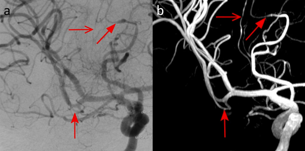
Biagi L, Lenzi S, Cipriano E, Fiori S, Bosco P, Cristofani P, Astrea G, Pini A, Cioni G, Mercuri E, Tosetti M, Battini R
Neural substrates of neuropsychological profiles in dystrophynopathies: A pilot study of diffusion tractography imaging
Plos one, 16(5):e0250420, 2021

ABSTRACT: Introduction: Cognitive difficulties and neuropsychological alterations in Duchenne and Becker muscular dystrophy (DMD, BMD) boys are not yet sufficiently explored, although this topic could have a relevant impact, finding novel biomarkers of disease both at genetics and neuroimaging point of view. The current study aims to: 1) analyze the neuropsychological profile of a group of DMD and BMD boys without cognitive impairment with an assessment of their executive functions; 2) explore the structural connectivity in DMD, BMD, and age-matched controls focusing on cortico-subcortical tracts that connect frontal cortex, basal ganglia, and cerebellum via the thalamus; 3) explore possible correlations between altered structural connectivity and clinical neuropsychological measures.
Materials and methods: This pilot study included 15 boys (5 DMD subjects, 5 BMD subjects, and 5 age-matched typically developing, TD). They were assessed using a neuropsychological assessment protocol including cognitive and executive functioning assessment and performed a 1.5T MRI brain exam including advance Diffusion Weighted Imaging (DWI) method for tractography. Structural connectivity measurements were extracted along three specific tracts: Cortico-Ponto-Cerebellar Tract (CPCT), Cerebellar-Thalamic Tract (CTT), and Superior Longitudinal Fasciculus (SLF). Cortical-Spinal Tract (CST) was selected for reference, as control tract.
Results: Regarding intellectual functioning, a major impairment in executive functions compared to the general intellectual functioning was observed both for DMD (mean score = 86.20; SD = 11.54) and for BMD children (mean score = 88; SD = 3.67). Mean FA resulted tendentially always lower in DMD compared to both BMD and TD groups for all the examined tracts. The differences in FA were statistically significant for the right CTT (DMD vs BMD, p = 0.002, and DMD vs TD, p = 0.0015) and the right CPCT (DMD vs TD, p = 0.008). Concerning DMD, significant correlations emerged between FA-R-CTT and intellectual quotients (FIQ, p = 0.044; ρs = 0.821), and executive functions (Denomination Total, p = 0.044, ρs = 0.821; Inhibition Total, p = 0.019, ρs = 0.900). BMD showed a significant correlation between FA-R-CPCT and working memory index (p = 0.007; ρs = 0.949).
Discussion and conclusion: In this pilot study, despite the limitation of sample size, the findings support the hypothesis of the involvement of a cerebellar-thalamo-cortical loop for the neuropsychological profile of DMD, as the CTT and the CPCT are involved in the network and the related brain structures are known to be implied in executive functions. Our results suggest that altered WM connectivity and reduced fibre organization in cerebellar tracts, probably due to the lack of dystrophin in the brain, may render less efficient some neuropsychological functions in children affected by dystrophinopathies. The wider multicentric study could help to better establish the role of cerebellar connectivity in neuropsychological profile for dystrophinopathies, identifying possible novel diagnostic and prognostic biomarkers.
Shridhar Konar A, Qian E, Geethanath S, Buonincontri G, Obuchowski NA, Fung M, Gomez P, Schulte R, Cencini M, Tosetti M, Schwartz LH, Shukla‐Dave A
Quantitative imaging metrics derived from magnetic resonance fingerprinting using ISMRM/NIST MRI system phantom: An international multicenter repeatability and reproducibility study
Plos one, 16(5):e0250420, 2021

ABSTRACT: Purpose: To compare the bias and inherent reliability of the quantitative (T1 and T2) imaging metrics generated from the magnetic resonance fingerprinting (MRF) technique using the ISMRM/NIST system phantom in an international multicenter setting.
Method: ISMRM/NIST MRI system phantom provides standard reference T1 and T2 relaxation values (vendor-provided) for each of the 14 vials in T1 and T2 arrays. MRF-SSFP scans repeated over 30 days on GE 1.5 and 3.0 T scanners at three collaborative centers. MRF estimated T1, and T2 values averaged over 30 days were compared with the phantom vendor-provided and spin-echo (SE) based convention gold standard (GS) method. Repeatability and reproducibility were characterized by the within-case coefficient of variation (wCV) of the MRF data acquired over 30 days, along with the biases.
Result: For the wide ranges of MRF estimated T1 values, vials #1-8 (T1 relaxation time between 2033 and 184 ms) exhibited a wCV less than 3% and 4%, respectively, on 3.0 and 1.5 T scanners. T2 values in vials #1-8 (T2 relaxation, 1044-45 ms) have shown wCV to be <7% on both 3.0 and 1.5 T scanners. A stronger linear correlation overall for T1 (R2 = 0.9960 and 0.9963 at center-1 and center-2 on 3.0 T scanner, and R2 = 0.9951 and 0.9988 at center-1 and center-3 on 1.5 T scanner) compared to T2 (R2 = 0.9971 and 0.9972 at center-1 and center-2 on 3.0 T scanner, and R2 = 0.9815 and 0.9754 at center-1 and center-3 on 1.5 T scanner). Bland–Altman (BA) analysis showed MRF based T1 and T2 values were within the limit of agreement (LOA) except for one data point. The mean difference or bias and 95% lower bound (LB) and upper bound (UB) LOA are reported in the format; mean bias: 95% LB LOA: 95% UB LOA. The biases for T1 values were 21.34: −50.00: 92.69, 21.32: −47.29: 89.94 ms, and for T2 values were −19.88: −42.37: 2.61, −19.06: −43.58: 5.45 ms on 3.0 T scanner at center-1 and center-2, respectively. Similarly, on 1.5 T scanner biases for T1 values were 26.54: −53.41: 106.50, 9.997: −51.94: 71.94 ms, and for T2 values were −23.84: −135.40: 87.76, −37.30: 134.30: 59.73 ms at center-1 and center-3, respectively. Additionally, the correlation between the SE based GS and MRF estimated T1 and T2 values (R2 = 0.9969 and 0.9977) showed a similar trend as we observed between vendor-provided and MRF estimated T1 and T2 values (R2 = 0.9963 and 0.9972). In addition to correlation, BA analysis showed that all the vials are within the LOA between the GS and vendor-provided for the T1 values and except one vial for T2. All the vials are within the LOA between GS and MRF except one vial in T1 and T2 array. The wCV for reproducibility was <3% for both T1 and T2 values in vials #1-8, for all the 14 vials, wCV calculated for reproducibility was <4% for T1 values and <5% for T2.
Conclusion: This study shows that MRF is highly repeatable (wCV <4% for T1 and <7% for T2) and reproducible (wCV < 3% for both T1 and T2) in certain vials (vials #1-8).
Buonincontri G, Kurzawski JW, Kaggie JD, Matys T, Gallagher FA, Cencini M, Donatelli G, Cecchi P, Cosottini M, Martini N, Frijia F, Montanaro D, Gómez PA, Schulte RF, Retico A, Tosetti M
Three dimensional MRF obtains highly repeatable and reproducible multi-parametric estimations in the healthy human brain at 1.5T and 3T
NeuroImage, 226:117573, 2021

ABSTRACT: Magnetic resonance fingerprinting (MRF) is highly promising as a quantitative MRI technique due to its accuracy, robustness, and efficiency. Previous studies have found high repeatability and reproducibility of 2D MRF acquisitions in the brain. Here, we have extended our investigations to 3D MRF acquisitions covering the whole brain using spiral projection k-space trajectories. Our travelling head study acquired test/retest data from the brains of 12 healthy volunteers and 8 MRI systems (3 systems at 3 T and 5 at 1.5 T, all from a single vendor), using a study design not requiring all subjects to be scanned at all sites. The pulse sequence and reconstruction algorithm were the same for all acquisitions. After registration of the MRF-derived PD T1 and T2 maps to an anatomical atlas, coefficients of variation (CVs) were computed to assess test/retest repeatability and inter-site reproducibility in each voxel, while a General Linear Model (GLM) was used to determine the voxel-wise variability between all confounders, which included test/retest, subject, field strength and site. Our analysis demonstrated a high repeatability (CVs 0.7–1.3% for T1, 2.0–7.8% for T2, 1.4–2.5% for normalized PD) and reproducibility (CVs of 2.0–5.8% for T1, 7.4–10.2% for T2, 5.2–9.2% for normalized PD) in gray and white matter. Both repeatability and reproducibility improved when compared to similar experiments using 2D acquisitions. Three-dimensional MRF obtains highly repeatable and reproducible estimations of T1 and T2, supporting the translation of MRF-based fast quantitative imaging into clinical applications.
2020
Conti E, Retico A, Palumbo L, Spera G, Bosco P, Biagi L, Fiori S, Tosetti M, Cipriani P, Cioni G, Muratori F, Chilosi A, Calderoni S
Autism spectrum disorder and childhood apraxia of speech: early language-related hallmarks across structural MRI study
NeuroImage, 226:117573, 2020

ABSTRACT: Autism Spectrum Disorder (ASD) and Childhood Apraxia of Speech (CAS) are developmental disorders with distinct diagnostic criteria and different epidemiology. However, a common genetic background as well as overlapping clinical features between ASD and CAS have been recently reported. To date, brain structural language-related abnormalities have been detected in both the conditions, but no study directly compared young children with ASD, CAS and typical development (TD). In the current work, we aim: (i) to test the hypothesis that ASD and CAS display neurostructural differences in comparison with TD through morphometric Magnetic Resonance Imaging (MRI)-based measures (ASD vs. TD and CAS vs. TD); (ii) to investigate early possible disease-specific brain structural patterns in the two clinical groups (ASD vs. CAS); (iii) to evaluate predictive power of machine-learning (ML) techniques in differentiating the three samples (ASD, CAS, TD). We retrospectively analyzed the T1-weighted brain MRI scans of 68 children (age range: 34–74 months) grouped into three cohorts: (1) 26 children with ASD (mean age ± standard deviation: 56 ± 11 months); (2) 24 children with CAS (57 ± 10 months); (3) 18 children with TD (55 ± 13 months). Furthermore, a ML analysis based on a linear-kernel Support Vector Machine (SVM) was performed. All but one brain structures displayed significant higher volumes in both ASD and CAS children than TD peers. Specifically, ASD alterations involved fronto-temporal regions together with basal ganglia and cerebellum, while CAS alterations are more focused and shifted to frontal regions, suggesting a possible speech-related anomalies distribution. Caudate, superior temporal and hippocampus volumes directly distinguished the two conditions in terms of greater values in ASD compared to CAS. The ML analysis identified significant differences in brain features between ASD and TD children, whereas only some trends in the ML classification capability were detected in CAS as compared to TD peers. Similarly, the MRI structural underpinnings of two clinical groups were not significantly different when evaluated with linear-kernel SVM. Our results may represent the first step towards understanding shared and specific neural substrate in ASD and CAS conditions, which subsequently may contribute to early differential diagnosis and tailoring specific early intervention.
Cervelli R, Cencini M, Buonincontri G, Campana F, Cacciato Insilla A, Aringhieri G, De Simone P, Boggi U, Campani D, Tosetti M, Crocetti L
7-T MRI of explanted liver and ex-vivo pancreatic specimens: prospective study protocol of radiological-pathological correlation feasibility (the EXLIPSE project)
European radiology experimental, 4(1):58, 2020

ABSTRACT: The study focuses on radiological-pathological correlation between imaging of ex vivo samples obtained by a 7-T scanner and histological examination. The specimens will be derived from native explanted cirrhotic livers, liver grafts excluded from donation because of severe steatosis, and primary pancreatic tumours. Magnetic resonance imaging (MRI) examinations will be performed within 24 h from liver or pancreatic lesion surgical removal. The MRI protocol will include morphological sequences, quantitative T1, T2, and fat-, water-fraction maps with Cartesian k-space acquisition, and multiparametric methods based on a transient-state “MRI fingerprinting”. Finally, the specimen will be fixed by formalin. Qualitative imaging analysis will be performed by two independent blinded radiologists to assess image consistency score. Quantitative analysis will be performed by drawing regions of interest on different tissue zones to measure T1 and T2 relaxation times as well as fat- and water-fraction. The same tissue areas will be analysed by the pathologists. This study will provide the possibility to improve our knowledge about qualitative and quantitative abdominal imaging assessment at 7 T, by correlating imaging characteristics and the corresponding histological composition of ex vivo specimens, in order to identify imaging biomarkers.
Aringhieri G, Zampa V, Tosetti M
Musculoskeletal MRI at 7 T: do we need more or is it more than enough?
European radiology experimental, 4(1):48, 2020

ABSTRACT: Ultra-high field magnetic resonance imaging (UHF-MRI) provides important diagnostic improvements in musculoskeletal imaging. The higher signal-to-noise ratio leads to higher spatial and temporal resolution which results in improved anatomic detail and higher diagnostic confidence. Several methods, such as T2, T2*, T1rho mapping, delayed gadolinium-enhanced, diffusion, chemical exchange saturation transfer, and magnetisation transfer techniques, permit a better tissue characterisation. Furthermore, UHF-MRI enables in vivo measurements by low-γ nuclei (23Na, 31P, 13C, and 39K) and the evaluation of different tissue metabolic pathways. European Union and Food and Drug Administration approvals for clinical imaging at UHF have been the first step towards a more routinely use of this technology, but some drawbacks are still present limiting its widespread clinical application. This review aims to provide a clinically oriented overview about the application of UHF-MRI in the different anatomical districts and tissues of musculoskeletal system and its pros and cons. Further studies are needed to consolidate the added value of the use of UHF-MRI in the routine clinical practice and promising efforts in technology development are already in progress.
Kurzawski JW, Cencini M, Peretti L, Gómez PA, Schulte RF, Donatelli G, Cosottini M, Cecchi P, Costagli M, Retico A, Tosetti M, Buonincontri G
Retrospective rigid motion correction of three‐dimensional magnetic resonance fingerprinting of the human brain
Magnetic resonance in medicine, 84(5):2606-2615, 2020

ABSTRACT: Purpose: To obtain three-dimensional (3D), quantitative and motion-robust imaging with magnetic resonance fingerprinting (MRF).
Methods: Our acquisition is based on a 3D spiral projection k-space scheme. We compared different orderings of trajectory interleaves in terms of rigid motion-correction robustness. In all tested orderings, we considered the whole dataset as a sum of 56 segments of 7-s duration, acquired sequentially with the same flip angle schedule. We performed a separate image reconstruction for each segment, producing whole-brain navigators that were aligned to the first segment using normalized correlation. The estimated rigid motion was used to correct the k-space data, and the aligned data were matched with the dictionary to obtain motion-corrected maps.
Results: A significant improvement on the motion-affected maps after motion correction is evident with the suppression of motion artifacts. Correlation with the motionless baseline improved by 20% on average for both T1 and T2 estimations after motion correction. In addition, the average motion-induced quantification bias of 70 ms for T1 and 18 ms for T2 values was reduced to 12 ms and 6 ms, respectively, improving the reliability of quantitative estimations.
Conclusion: We established a method that allows correcting 3D rigid motion on a 7-s timescale during the reconstruction of MRF data using self-navigators, improving the image quality and the quantification robustness.
Sgandurra G, Biagi L, Fogassi L, Ferrari A, Sicola E, Guzzetta A, Tosetti M, Cioni G
Reorganization of action observation and sensory-motor networks after action observation therapy in children with congenital hemiplegia: A pilot study
Developmental Neurobiology, 80(9-10):351-360, 2020

ABSTRACT: New rehabilitation programs based on action observation therapy (AOT) are effective in improving motor function in children with congenital hemiplegia. In this pilot study we tested the potential effects of AOT on the reorganization of the motor system by functional magnetic resonance imaging (fMRI). As part of a randomized trial, eight subjects (age range: 6.2–14.5 years) with congenital hemiplegia were randomly assigned to an experimental (EG) or control (CG) group. All children underwent a clinical and neurophysiological assessment with Assisting Hand Assessment (AHA), MRI, and fMRI at baseline (T0), 1(T1), and 8(T2) weeks after the end of 3-week treatment. For the EG, AOT consisted in the observation of uni/bimanual goal-directed actions followed by their execution. CG watched same-duration computer games and then performed the same actions in the same order used in the EG. fMRI study was carried out using two different paradigms, for exploring sensory-motor network (SMN) localization and action observation network (AON). The pattern of brain activation was generally similar between T0 and T1 for both groups, while it was more widespread at T2, compared to T0 and T1, in the EG. This enlargement was coupled with functional improvement at AHA. Single-subject analysis shows a reduction of lateralization indexes both for the AON and the SMN. This pilot study, despite the small sample, showed the fMRI feasibility for providing relevant biomarkers of brain plasticity for monitoring the AOT response in children with congenital hemiplegia.
Gómez PA, Cencini M, Golbabaee M, Schulte RF, Pirkl C, Horvath I, Fallo G, Peretti L, Tosetti M, Menze BH, Buonincontri G
Rapid three-dimensional multiparametric MRI with quantitative transient-state imaging
Scientific Reports, 10:13769, 2020
ABSTRACT: Novel methods for quantitative, transient-state multiparametric imaging are increasingly being demonstrated for assessment of disease and treatment efficacy. Here, we build on these by assessing the most common Non-Cartesian readout trajectories (2D/3D radials and spirals), demonstrating efficient anti-aliasing with a k-space view-sharing technique, and proposing novel methods for parameter inference with neural networks that incorporate the estimation of proton density. Our results show good agreement with gold standard and phantom references for all readout trajectories at 1.5 T and 3 T. Parameters inferred with the neural network were within 6.58% difference from the parameters inferred with a high-resolution dictionary. Concordance correlation coefficients were above 0.92 and the normalized root mean squared error ranged between 4.2 and 12.7% with respect to gold-standard phantom references for T1 and T2. In vivo acquisitions demonstrate sub-millimetric isotropic resolution in under five minutes with reconstruction and inference times < 7 min. Our 3D quantitative transient-state imaging approach could enable high-resolution multiparametric tissue quantification within clinically acceptable acquisition and reconstruction times.
2019
Düzel E, Acosta‐Cabronero J, Berron D, Biessels GJ, Björkman‐Burtscher I, Bottlaender M, Bowtell R, v Buchem M, Cardenas‐Blanco A, Boumezbeur F, Chan D, Clare S, Costagli M, de Rochefort L, Fillmer A, Gowland P, Hansson O, Hendrikse J, Kraff O, Ladd ME, Ronen I, Petersen E, Rowe JB, Siebner H, Stoecker T, Straub S, Tosetti M, Uludag K, Vignaud A, Zwanenburg J, Speck O
European Ultrahigh-Field Imaging Network for Neurodegenerative Diseases (EUFIND)
Alzheimer’s & Dementia: Diagnosis, Assessment & Disease Monitoring, 197, 557-564, 2019

ABSTRACT: Introduction: The goal of European Ultrahigh-Field Imaging Network in Neurodegenerative Diseases (EUFIND) is to identify opportunities and challenges of 7 Tesla (7T) MRI for clinical and research applications in neurodegeneration. EUFIND comprises 22 European and one US site, including over 50 MRI and dementia experts as well as neuroscientists.
Methods: EUFIND combined consensus workshops and data sharing for multisite analysis, focusing on 7 core topics: clinical applications/clinical research, highest resolution anatomy, functional imaging, vascular systems/vascular pathology, iron mapping and neuropathology detection, spectroscopy, and quality assurance. Across these topics, EUFIND considered standard operating procedures, safety, and multivendor harmonization.
Results: The clinical and research opportunities and challenges of 7T MRI in each subtopic are set out as a roadmap. Specific MRI sequences for each subtopic were implemented in a pilot study presented in this report. Results show that a large multisite 7T imaging network with highly advanced and harmonized imaging sequences is feasible and may enable future multicentre ultrahigh-field MRI studies and clinical trials.
Discussion: The EUFIND network can be a major driver for advancing clinical neuroimaging research using 7T and for identifying use-cases for clinical applications in neurodegeneration.
Lancione M, Donatelli G, Cecchi P, Cosottini M, Tosetti M, Costagli M
Echo time dependency of quantitative susceptibility mapping reproducibility at different magnetic field strength
NeuroImage, 197, 557-564, 2019

ABSTRACT: Quantitative Susceptibility Mapping (QSM) provides a way of measuring iron concentration and myelination non-invasively and has the potential of becoming a tool of paramount importance in the study of a host of different pathologies. However, several experimental factors and the physical properties of magnetic susceptibility (χ) can impair the reliability of QSM, and it is therefore essential to assess QSM reproducibility for repeated acquisitions and different field strength. In particular, it has recently been demonstrated that QSM measurements strongly depend on echo time (TE): the same tissue, measured on the same scanner, exhibits different apparent frequency shifts depending on the TE used. This study aims to assess the influence of TE on intra-scanner and inter-scanner reproducibility of QSM, by using MRI systems operating at 3T and 7T. To maximize intra-scanner reproducibility it is necessary to match the TEs of the acquisition protocol, but the application of this rule leads to inconsistent QSM values across scanners operating at different static magnetic field. This study however demonstrates that, provided a careful choice of acquisition parameters, and in particular by using TEs at 3T that are approximately 2.6 times longer than those at 7T, highly reproducible whole-brain χ maps can be achieved also across different scanners, which renders QSM a suitable technique for longitudinal follow-up in clinical settings and in multi-center studies.
Buonincontri G, Biagi L, Retico A, Cecchi P, Cosottini M, Gallagher FA, Gómez PA, Graves MJ, McLean MA, Riemer F, Schulte RF, Tosetti M, Zaccagna F, Kaggie JD
Multi-site repeatability and reproducibility of MR fingerprinting of the healthy brain at 1.5 and 3.0 T
NeuroImage, 195, 362-372, 2019

ABSTRACT: Fully-quantitative MR imaging methods are useful for longitudinal characterization of disease and assessment of treatment efficacy. However, current quantitative MRI protocols have not been widely adopted in the clinic, mostly due to lengthy scan times. Magnetic Resonance Fingerprinting (MRF) is a new technique that can reconstruct multiple parametric maps from a single fast acquisition in the transient state of the MR signal. Due to the relative novelty of this technique, the repeatability and reproducibility of quantitative measurements obtained using MRF has not been extensively studied. Our study acquired test/retest data from the brains of nine healthy volunteers, each scanned on five MRI systems (two at 3.0 T and three at 1.5 T, all from a single vendor) located at two different centers. The pulse sequence and reconstruction algorithm were the same for all acquisitions. After registration of the MRF-derived M0, T1 and T2 maps to an anatomical atlas, coefficients-of-variation (CVs) were computed to assess test/retest repeatability and inter-site reproducibility in each voxel, while a General Linear Model (GLM) was used to determine the voxel-wise variability between all confounders, which included test/retest, subject, field strength and site. Our analysis demonstrated an excellent repeatability (CVs of 2–3% for T1, 5–8% for T2, 3% for normalized-M0) and a good reproducibility (CVs of 3–8% for T1, 8–14% for T2, 5% for normalized-M0) in grey and white matter.
Gagliardi V, Puccini A, Belcari N, Carmarlinghi N, Fantacci ME, Fidecaro F, Franchi G, Sportelli G, Biagi L, Tosetti M, Bisogni MG
MR Compatible Power Supply Module for PET Detectors of an Integrated PET/MR System
IEEE Transactions on Radiation and Plasma Medical Sciences, 3(4):454-464, 2019
ABSTRACT: Power supply of positron emission tomography (PET) detectors is a serious issue for PET/magnetic resonance imaging (MRI) systems, due to mutual interaction between power supply electronics and MRI magnetic fields. We developed a prototype of a magnetic compatible power supply, that can be placed in an MRI system. The power supply is a compact (5.2×5.2 cm 2 ) iron-free switching dc-dc converter. The power supply works in the very high frequency (VHF) range, between 24.5 and 26.5 MHz, to allow using air core MR compatible inductors, and avoiding RF coupling between harmonics of power supply signal and RF magnetic field of a 1.5 T MRI scanner. The power supply has been designed as an upgrades for the TRIMAGE system and is able to convert 12-V dc input to 5-V dc output with a measured efficiency of 71.43%. Magnetic compatibility has been verified in the 1.5 T magnet of the IRCCS Stella Maris Foundation, Pisa, Italy, and in the 7 T MRI of the IMAGO7 Foundation, Pisa, Italy. Performance measurements of the power supply showed no variations during the magnetic compatibility tests. MRI scanner performance was similar with and without the power supply, and no artifacts were induced by the circuit in the acquired MRI images.
Maggiorelli F, Boskamp EB, Tiberi G, Retico A, Symms MR, Tosetti M, Robb F
Double-Tuned Surface 1H–23Na Radio Frequency Coils at 7 T: Comparison of Three Decoupling Methods
Applied Magnetic Resonance, 50(5):649-661, 2019
ABSTRACT: Magnetic Resonance Imaging (MRI) and Spectroscopy (MRS) with nuclei other than protons (X-nuclei) often require the acquisition of proton signal for shimming and co-registration procedures. Double-Tuned Radio Frequency (DT-RF) coils improve these procedures, avoiding the need for movement and repositioning of the subject during the examination. The drawback of DT-RF coils is basically the coupling between the two resonant structures, which increases signal losses leading to a degradation of the final MR image. To improve MR signal quality acquired via DT-RF coils, a suitable decoupling strategy should be implemented. For this purpose, three DT-RF coil prototypes, which differed only in the decoupling method, were built and their performances were compared through workbench measurements. Each prototype consisted of two concentric loops. The inner and outer loops were tuned at sodium (≈ 79 MHz) and proton (≈ 300 MHz) Larmor frequency at 7 Tesla, respectively. Active and passive decoupling designs were compared measuring the Q factor and the S21 parameter for each prototype. Active decoupling was tested as an alternative to the standard passive decoupling with a trap circuit, in which a non-negligible amount of current flows at resonance, perturbing the magnetic field responsible for producing the MR image. Workbench measurements showed satisfactory Q factors and S21 for both active and passive decoupling cases. Thus, active decoupling could be a promising alternative to achieve better MR signal quality. Furthermore, for active decoupling, two circuit elements were examined: PIN diodes and micro-electromechanical system (MEMS) switches.
Cencini M, Biagi L, Kaggie JD, Schulte RF, Tosetti M, Buonincontri G
Magnetic resonance fingerprinting with dictionary‐based fat and water separation (DBFW MRF): a multi‐component approach
Magnetic resonance in medicine, 81(5):3032-3045, 2019

ABSTRACT: Purpose: To obtain a fast and robust fat-water separation with simultaneous estimation of water T1, fat T1, and fat fraction maps.
Methods: We modified an MR fingerprinting (MRF) framework to use a single dictionary combination of a water and fat dictionary. A variable TE acquisition pattern with maximum TE = 4.8 ms was used to increase the fat–water separability. Radiofrequency (RF) spoiling was used to reduce the size of the dictionary by reducing T2 sensitivity. The technique was compared both in vitro and in vivo to an MRF method that incorporated 3-point Dixon (DIXON MRF), as well as Cartesian IDEAL with different acquisition parameters.
Results: The proposed dictionary-based fat–water separation technique (DBFW MRF) successfully provided fat fraction, water, and fat T1, B0, and B1+ maps both in vitro and in vivo. The fat fraction and water T1 values obtained with DBFW MRF show excellent agreement with DIXON MRF as well as with the reference values obtained using a Cartesian IDEAL with a long TR (concordance correlation coefficient: 0.97/0.99 for fat fraction–water T1). Whereas fat fraction values with Cartesian IDEAL were degraded in the presence of T1 saturation, MRF methods successfully estimated and accounted for T1 in the fat fraction estimates.
Conclusion: The DBFW MRF technique can successfully provide T1 and fat fraction quantification in under 20 s per slice, intrinsically correcting T1 biases typical of fast Dixon techniques. These features could improve the diagnostic quality and use of images in presence of fat.
Cencini M, Tosetti M, Buonincontri G
An aristotelian view on MR-based attenuation correction (ARISTOMRAC): Combining the four elements
IEEE Transactions on Radiation and Plasma Medical Sciences, 3(4):491-497, 2019

ABSTRACT: Magnetic resonance-based attenuation correction (MRAC) is important for accurate quantification of the uptake of PET tracers in combined PET/MR scanners. However, current techniques for MRAC usually require multiple acquisitions or complex post-processing to discriminate the different tissues. Inspired by the ancient Greeks, who believed that matter was made of the combination of four elements (earth, water, air, and fire), we formulated a multicomponent magnetic resonance (MR) fingerprinting framework, where every voxel was considered a weighted combination of four base elements: 1) bone; 2) water; 3) air; and 4) fat. We named our approach aristotelian MR-based attenuation correction (ARISTOMRAC). We used a 3-D radial acquisition scheme at 1.5T, acquiring a transient-state spoiled acquisition with variable flip angles and echo times (TE), with the shortest TEs being ultrashort echo times (UTEs). We simulated a multitissue MR signal model using the Bloch equations and used dictionary matching to extract tissue fraction maps for bone water and fat, while air fractions were obtained by thresholding the UTE parts of our acquisitions at higher spatial resolution. Compared to previous methods for MRAC, our approach used a full multicomponent signal model, including multiple tissues per voxel. For this reason, rather than reconstructing high resolutions images, MR data can be acquired more efficiently, directly at the resolution needed for PET attenuation maps. The ARISTOMRAC method allows to accurately estimate the air, water, bone, and fat fractions (concordance correlation coefficient = 0.81/0.91/0.98 for bone, water, and fat, respectively). Attenuation maps could be obtained in the head and neck with a single 1-min acquisition.
Chilosi AM, Bulgheroni S, Turi M, Cristofani P, Biagi L, Erbetta A, Riva D, Nigri A, Caputi L, Giannini N, Fiori S, Pecini C, Perego F, Tosetti M, Cipriani P, Cioni G
Hemispheric language organization after congenital left brain lesions: a comparison between functional transcranial Doppler and functional MRI
Journal of Neuropsychology, 13(1):46-66, 2019
ABSTRACT: This study investigated whether functional transcranial Doppler ultrasound (fTCD) is a suitable tool for studying hemispheric lateralization of language in patients with pre-perinatal left hemisphere (LH) lesions and right hemiparesis. Eighteen left-hemisphere-damaged children and young adults and 18 healthy controls were assessed by fTCD and fMRI to evaluate hemispheric activation during two language tasks: a fTCD animation description task and a fMRI covert rhyme generation task. Lateralization indices (LIs), measured by the two methods, differed significantly between the two groups, for a clear LH dominance in healthy participants and a prevalent activation of right hemisphere in more than 80% of brain-damaged patients. Distribution of participants in terms of left, right, and bilateral lateralization was highly concordant between fTCD and fMRI values. Moreover, right hemisphere language dominance in patients with left hemispheric lesions was significantly associated with severity of cortical and subcortical damage in LH. This study suggests that fTCD is an easily applicable tool that might be a valid alternative to fMRI for large-scale studies of patients with congenital brain lesions.
Costagli M, Lancione M, Cecchetti L, Pietrini P, Cosottini M, Ricciardi E, Tosetti M
Quantitative Susceptibility Mapping of Brain Function during Auditory Stimulation
IEEE Transactions on Radiation and Plasma Medical Sciences, 2019

ABSTRACT: Functional quantitative susceptibility mapping (fQSM) is a recently established method that, based on the same acquisition technique as conventional functional magnetic resonance imaging, has two very appealing features: 1) it is quantitative and 2) it is considerably less affected by nonlocal effects than the blood oxygenation level-dependent (BOLD) signal. Here, the response of the auditory cortex to the presentation of short acoustic stimuli has been studied with fQSM. fQSM responses to stimuli were obtained by using a data-driven approach based on signal deconvolution to avoid assumptions regarding the response shape. fQSM and BOLD responses showed similar shapes, however, while the majority (82%) of fQSM responses described transient decreases in magnetic susceptibility (explainable by the same mechanisms behind the typical, positive BOLD responses), some voxels exhibited transient increments in magnetic susceptibility, which might reflect the interplay among changes in fractional oxygen saturation, cerebral blood flow, and volume. Statistically significant fQSM responses were observed not only in veins (which constitute the largest sources of magnetic susceptibility contrast) but also in gray matter tissue, suggesting that this technique is sensitive to subtle, stimulus-dependent changes also in the parenchyma, and therefore it might be useful to localize and interpret the sources of activation.
2018
Giovannetti G, Tiberi G, Tosetti M, Monorchio A, Fontana N
Radiofrequency planar surface coil for magnetic resonance: When the use of a circular wire gives a noticeable advantage with respect to a flat strip conductor?
Measurement, 129:518-522, 2018
ABSTRACT: Depending on their cross-sectional shape, commonly used conductors for radiofrequency (RF) Magnetic Resonance (MR) coils can be categorized into circular wires and flat strips. Due to a more symmetrical current distribution inside conductor volume, coils constituted by wire conductors provide better overall performance in unloaded conditions with respect to the ones made of strip conductor, although wire conductors are difficult to handle for coil manufacturing and additional mechanical competencies are required. Nevertheless, the accomplishment of the best coil performance during imaging, i.e. in the presence of a sample, remains the main issue in MRI. It follows that the use of wire conductors instead of strip ones is worthwhile only if the correspondent increase in coil quality factor with sample is substantial: this is related to the ratio between sample and coil resistance. This paper proposes the application of a finite element method (FEM)-based numerical approach for separately estimating the conductor and radiative losses in planar surface loops characterized by different cross-sectional shapes (circular wire and flat strip) in conjunction with a vector potential calculation-based method for sample-induced resistance estimation. Simulation data were acquired from 5.7 to 128 MHz, for four different size loops (from 2 to 15 cm diameters), with the scope of evaluating the region in the frequency-loop diameter plane where the use of a circular wire conductor gives a noticeable advantage with respect to the flat strip in maximizing signal-to-noise ratio (SNR) in MR applications.
Binda P, Kurzawski JW, Lunghi C, Biagi L, Tosetti M, Morrone MC
Response to short-term deprivation of the human adult visual cortex measured with 7T BOLD
eLife, 7:e40014, 2018
ABSTRACT: Sensory deprivation during the post-natal ‘critical period’ leads to structural reorganization of the developing visual cortex. In adulthood, the visual cortex retains some flexibility and adapts to sensory deprivation. Here we show that short-term (2 hr) monocular deprivation in adult humans boosts the BOLD response to the deprived eye, changing ocular dominance of V1 vertices, consistent with homeostatic plasticity. The boost is strongest in V1, present in V2, V3 and V4 but absent in V3a and hMT+. Assessment of spatial frequency tuning in V1 by a population Receptive-Field technique shows that deprivation primarily boosts high spatial frequencies, consistent with a primary involvement of the parvocellular pathway. Crucially, the V1 deprivation effect correlates across participants with the perceptual increase of the deprived eye dominance assessed with binocular rivalry, suggesting a common origin. Our results demonstrate that visual cortex, particularly the ventral pathway, retains a high potential for homeostatic plasticity in the human adult.
Fiori S, Biagi L, Cecchi C, Cioni G, Beani E, Tosetti M, Cosottini M, Guzzetta G
Potentials of ultrahigh-field MRI for the study of somatosensory reorganization in congenital hemiplegia
Neural plasticity, 2018:8472807, 2018
ABSTRACT: Reorganization of somatosensory function influences the clinical recovery of subjects with congenital unilateral brain lesions. Ultrahigh-field (UHF) functional MRI (fMRI) with the use of a 7 T magnet has the potential to contribute fundamentally to the current knowledge of such plasticity mechanisms. The purpose of this study was to obtain preliminary information on the possible advantages of the study of somatosensory reorganization at UHF fMRI. We enrolled 6 young adults (mean age 25 ± 6 years) with congenital unilateral brain lesions (4 in the left hemisphere and 2 in the right hemisphere; 4 with perilesional motor reorganization and 2 with contralesional motor reorganization) and 7 healthy age-matched controls. Nondominant hand sensory assessment included stereognosis and 2-point discrimination. Task-dependent fMRI was performed to elicit a somatosensory activation by using a safe and quantitative device developed ad hoc to deliver a reproducible gentle tactile stimulus to the distal phalanx of thumb and index fingers. Group analysis was performed in the control group. Individual analyses in the native space were performed with data of hemiplegic subjects. The gentle tactile stimulus showed great accuracy in determining somatosensory cortex activation. Single-subject gentle tactile stimulus showed an S1 activation in the postcentral gyrus and an S2 activation in the inferior parietal insular cortex. A correlation emerged between an index of S1 reorganization (distance between expected and reorganized S1) and sensory deficit () in subjects with hemiplegia, with higher distance related to a more severe sensory deficit. Increase in spatial resolution at 7 T allows a better localization of reorganized tactile function validated by its correlation with clinical measures. Our results support the S1 early-determination hypothesis and support the central role of topography of reorganized S1 compared to a less relevant S1-M1 integration.
Sgandurra G, Biagi L, Fogassi L, Sicola E, Ferrari A, Guzzetta A, Tosetti M, Cioni G
Reorganization of the action observation network and sensory-motor system in children with unilateral cerebral palsy: an fMRI study
Neural plasticity, 2018:6950547, 2018
ABSTRACT: Little is known about the action observation network (AON) in children with unilateral cerebral palsy (UCP). Using fMRI, we aimed to explore AON and sensory-motor network (SMN) in UCP children and compare them to typically developed (TD) children and analyse the relationship between AON (re-)organization and several neurophysiological and clinical measures. Twelve UCP children were assessed with clinical scales and transcranial magnetic stimulation (TMS). For the fMRI study, they underwent a paradigm based on observation of complex and simple object-manipulation tasks executed by dominant and nondominant hand. Moreover, UCP and TD children carried out a further fMRI session to explore SMN in both an active motor and passive sensory task. AON in the UCP group showed higher lateralization, negatively related to performances on clinical scales, and had greater activation of unaffected hemisphere as compared to the bilateral representation in the TD group. In addition, a good congruence was found between bilateral or contralateral activation of AON and activation of SMN and TMS data. These findings indicate that our paradigm might be useful in exploring AON and the response to therapy in UCP subjects.
Donatelli G, Ceravolo R, Frosini D, Tosetti M, Bonuccelli U, Cosottini M
Present and Future of Ultra-High Field MRI in Neurodegenerative Disorders
Current Neurology and Neuroscience Reports, 18(6):1-15, 2018
ABSTRACT: Purpose of Review: With a high signal-to-noise ratio, unparalleled spatial resolution, and improved contrasts, ultra-high field MR (≥ 7 T) has great potential in depicting the normal radiological anatomy of smaller structures in the brain and can also provide more information about morphological, quantitative, and metabolic changes associated with a wide range of brain disorders. By focusing attention on specific brain regions believed to be associated with early pathological change, or by more closely inspecting recognized foci of brain pathology, ultra-high field MR can improve the accuracy and sensitivity of neuroimaging. This article reviews recent studies at ultra-high field about Alzheimer’s disease (AD), Parkinson’s disease (PD), and amyotrophic lateral sclerosis (ALS).
Recent Findings: The research on AD has mainly focused on detecting the thinning of hippocampal layers and the susceptibility effect supposed to be related to beta-amyloid deposition. In patients with PD, atypical parkinsonisms and subjects at risk of developing motor symptoms of Parkinson’s disease, the main aim was to detect changes in the substantia nigra, probably related to increased iron deposition. In patients with ALS, both brain and spinal cord were investigated, with the aim of finding changes in the primary motor cortex and corticospinal tract which reflect neurodegeneration and neuroinflammation.
Summary: Ultra-high field MR was shown to be useful for detecting subtle brain changes in patients with AD, and possible new diagnostic biomarkers in patients with PD and ALS were discovered. The ability of 7 T MR to provide prognostic biomarkers in subjects at risk for developing synucleinopathies is currently under evaluation.
Rua C, Wastling SJ, Costagli M, Symms MR, Biagi L, Cosottini M, Del Guerra A, Tosetti M, Barker GJ
Improving fMRI in signal drop-out regions at 7 T by using tailored radio-frequency pulses: application to the ventral occipito-temporal cortex
Magnetic Resonance Materials in Physics, Biology and Medicine, 31:257–267, 2018
ABSTRACT: Objective: Signal drop-off occurs in echo-planar imaging in inferior brain areas due to field gradients from susceptibility differences between air and tissue. Tailored-RF pulses based on a hyperbolic secant (HS) have been shown to partially recover signal at 3 T, but have not been tested at higher fields.
Materials and methods: The aim of this study was to compare the performance of an optimized tailored-RF gradient-echo echo-planar imaging (TRF GRE-EPI) sequence with standard GRE-EPI at 7 T, in a passive viewing of faces or objects fMRI paradigm in healthy subjects.
Results: Increased temporal-SNR (tSNR) was observed in the middle and inferior temporal lobes and orbitofrontal cortex of all subjects scanned, but elsewhere tSNR decreased relative to the standard acquisition. In the TRF GRE-EPI, increased functional signal was observed in the fusiform, lateral occipital cortex, and occipital pole, regions known to be part of the visual pathway involved in face-object perception.
Conclusion: This work highlights the potential of TRF approaches at 7 T. Paired with a reversed-gradient distortion correction to compensate for in-plane susceptibility gradients, it provides an improved acquisition strategy for future neurocognitive studies at ultra-high field imaging in areas suffering from static magnetic field inhomogeneities.
Donatelli G, Retico A, Caldarazzo Ienco E, Cecchi P, Costagli M, Frosini D, Biagi L, Tosetti M, Siciliano G, Cosottini M
Semiautomated evaluation of the primary motor cortex in patients with amyotrophic lateral sclerosis at 3T
American Journal of Neuroradiology, 39(1):63-69, 2018
ABSTRACT: Background and purpose: Amyotrophic lateral sclerosis is a neurodegenerative disease involving the upper and lower motor neurons. In amyotrophic lateral sclerosis, pathologic changes in the primary motor cortex include Betz cell depletion and the presence of reactive iron-loaded microglia, detectable on 7T MR images as atrophy and T2*-hypointensity. Our purposes were the following: 1) to investigate the signal hypointensity-to-thickness ratio of the primary motor cortex as a radiologic marker of upper motor neuron involvement in amyotrophic lateral sclerosis with a semiautomated method at 3T, 2) to compare 3T and 7T results, and 3) to evaluate whether semiautomated measurement outperforms visual image assessment.
Materials and methods: We investigated 27 patients and 13 healthy subjects at 3T, and 19 patients and 18 healthy subjects at 7T, performing a high-resolution 3D multiecho T2*-weighted sequence targeting the primary motor cortex. The signal hypointensity-to-thickness ratio of the primary motor cortex was calculated with a semiautomated method depicting signal intensity profiles of the cortex. Images were also visually classified as “pathologic” or “nonpathologic” based on the primary motor cortex signal intensity and thickness.
Results: The signal hypointensity-to-thickness ratio of the primary motor cortex was greater in patients than in controls (P < .001), and it correlated with upper motor neuron impairment in patients (ρ = 0.57, P < .001). The diagnostic accuracy of the signal hypointensity-to-thickness ratio was high at 3T (area under the curve = 0.89) and even higher at 7T (area under the curve = 0.94). The sensitivity of the semiautomated method (0.81) outperformed the sensitivity of the visual assessment (0.56–0.63) at 3T.
Conclusions: The signal hypointensity-to-thickness ratio of the primary motor cortex calculated with a semiautomated method is suggested as a radiologic marker of upper motor neuron burden in patients with amyotrophic lateral sclerosis. This semiautomated method may be useful for improving the subjective radiologic evaluation of upper motor neuron pathology in patients suspected of having amyotrophic lateral sclerosis.
2017
Lancione M, Tosetti M, Donatelli G, Cosottini M, Costagli M
The impact of white matter fiber orientation in single-acquisition quantitative susceptibility mapping
NMR in Biomedicine 30(11):e3798, 2017
ABSTRACT: The aim of this work was to assess the impact of tissue structural orientation on quantitative susceptibility mapping (QSM) reliability, and to provide a criterion to identify voxels in which measures of magnetic susceptibility (χ) are most affected by spatial orientation effects. Four healthy volunteers underwent 7-T magnetic resonance imaging (MRI). Multi-echo, gradient-echo sequences were used to obtain quantitative maps of frequency shift (FS) and χ. Information from diffusion tensor imaging (DTI) was used to investigate the relationship between tissue orientation and FS measures and QSM. After sorting voxels on the basis of their fractional anisotropy (FA), the variations in FS and χ values over tissue orientation were measured. Using a K-means clustering algorithm, voxels were separated into two groups depending on the variability of measures within each FA interval. The consistency of FS and QSM values, observed at low FA, was disrupted for FA > 0.6. The standard deviation of χ measured at high FA (0.0103 ppm) was nearly five times that at low FA (0.0022 ppm). This result was consistent through data across different head positions and for different brain regions considered separately, which confirmed that such behavior does not depend on structures with different bulk susceptibility oriented along particular angles. The reliability of single-orientation QSM anticorrelates with local FA. QSM provides replicable values with little variability in brain regions with FA < 0.6, but QSM should be interpreted cautiously in major and coherent fiber bundles, which are strongly affected by structural anisotropy and magnetic susceptibility anisotropy.
Andreuccetti D, Biagi L, Burriesci G, Cannatà V, Contessa GM, Falsaperla R, Genovese E, Lodato R, Lopresto V, Merla C, Napolitano A, Pinto R, Tiberi G, Tosetti N, Zoppetti N
Occupational exposure in MR facilities due to movements in the static magnetic field
Medical Physics, 44(11):5988-5996, 2017
ABSTRACT: Purpose: The exposure of operators moving in the static field of magnetic resonance (MR) facilities was assessed through measurements of the magnetic flux density, which is experienced as variable in time because of the movement. Collected data were processed to allow the comparison with most recent and authoritative safety standards.
Methods: Measurements of the experienced magnetic flux density B were performed using a probe worn by volunteers moving in MR environments. A total of 55 datasets were acquired nearby a 1.5 T, 3 T, and 7 T whole body scanners. Three different metrics were applied: the maximum intensity of B, to be compared with 2013/35/EU Directive exposure limit values for static fields; the maximum variation of the vector B on every 3s-interval, for comparison with the ICNIRP-2014 basic restriction aimed at preventing vertigo effects; two weighted-peak indices (for “sensory” and “health” effects: SENS-WP, HLTH-WP), assessing compliance with ICNIRP-2014 and EU Directive recommendations intended to prevent stimulation effects.
Results: Peak values of |B| were greater than 2 T in nine of the 55 datasets. All the datasets at 1.5 T and 3 T were compliant with the limit for vertigo effects, whereas six datasets at 7 T turned out to be noncompliant. At 7 T, all 36 datasets were noncompliant for the SENS-WP index and 26 datasets even for the HLTH-WP one.
Conclusions: Results demonstrate that compliance with EU Directive limits for static fields does not guarantee compliance with ICNIRP-2014 reference levels and clearly show that movements in the static field could be the key component of the occupational exposure to EMF in MR facilities.
Frosini D, Cosottini M, Donatelli G, Costagli M, Biagi L, Pacchetti C, Terzaghi M, Cortelli P, Arnaldi D, Bonanni E, Tosetti M, Bonuccelli U, Ceravolo R
Seven tesla MRI of the substantia nigra in patients with rapid eye movement sleep behavior disorder
Parkinsonism & related disorders, 43:105-109, 2017
ABSTRACT: Introduction: Susceptibility-weighted imaging of the substantia nigra (SN) both at 7 and 3 Tesla (T) has shown high accuracy in distinguishing patients with Parkinson’s disease (PD) and healthy subjects (HS). Patients with rapid eye movement (REM) behavior disorder (RBD) can develop synucleinopathies, and such risk is higher with dopamine transporter single photon emission tomography (123I-FP-CIT SPECT) evidence of nigro-striatal dysfunction. We aimed at evaluating SN 7T magnetic resonance imaging (7T-MRI) in patients with RBD and determining the agreement between MRI and 123I-FP-CIT SPECT.
Methods: Fifteen patients with idiopathic RBD confirmed by polysomnography and a recent 123I-FP-CIT SPECT underwent a 7T MR by using three-dimensional gradient-recalled-echo multiecho susceptibility-weighted imaging of the SN; the findings were randomly presented with those of 14 HS and 28 patients with PD and blindly evaluated by an expert neuroradiologist, according to recently published criteria. MRI and SPECT results were also compared.
Results: Nine subjects with RBD had abnormal SPECT; among them, the findings of 7T-MRI were rated abnormal in eight. Out of six subjects with RBD with normal SPECT, the 7T-MRI findings of five were rated normal. The Cohen’s kappa statistic value of agreement was 0.722.
Conclusion: Gradient-recalled-echo multiecho susceptibility-weighted imaging of the SN at 7T is abnormal in 60% of patients with RBD. The 7T-MRI and 123I-FP-CIT SPECT results showed good agreement. 7T-MRI of the SN could represent a safe marker for neurodegenerative disease in patients with RBD, however longitudinal study is warranted.
Buonincontri G, Schulte RF, Cosottini M, Tosetti M
Spiral MR fingerprinting at 7 T with simultaneous B1 estimation
Magnetic resonance imaging, 41:1-6, 2017
ABSTRACT: Magnetic resonance fingerprinting is an efficient, new approach for quantitative imaging with MR. We aimed to extend this technique to cases with B1 + inhomogeneities within the imaging volume.
Previous approaches have used abrupt changes in flip angles to estimate the B1 + field simultaneously with T1 and T2, using a Cartesian approach in a small-animal scanner at 4.7 T. Here, we evaluated whether a similar approach would be suitable for imaging human brains using spiral readouts with a 7 T scanner.
We found that our modified scheme could significantly reduce the adverse effects of B1 + inhomogeneities even in extreme cases, reducing both the bias and the variance in T2 estimations by an order of magnitude when compared to literature methods. Acquisitions used less than 1.5 W/kg SAR and could be performed in 12 s per slice.
In conclusion, our approach can be used to perform quantitative imaging of the brain at 7 T in a short time, simultaneously estimating the B1 + profile.
Retico A, Arezzini S, Bosco P, Calderoni S, Ciampa A, Coscetti S, Cuomo S, De Santis L, Fabiani D, Fantacci ME, Giuliano A, Mazzoni E, Mercatali P, Miscali G, Pardini M, Prosperi M, Romano F, Tamburini E, Tosetti M, Muratori F
ARIANNA: A research environment for neuroimaging studies in autism spectrum disorders
Computers in biology and medicine, 87:1-7, 2017
ABSTRACT: The complexity and heterogeneity of Autism Spectrum Disorders (ASD) require the implementation of dedicated analysis techniques to obtain the maximum from the interrelationship among many variables that describe affected individuals, spanning from clinical phenotypic characterization and genetic profile to structural and functional brain images. The ARIANNA project has developed a collaborative interdisciplinary research environment that is easily accessible to the community of researchers working on ASD (https://arianna.pi.infn.it). The main goals of the project are: to analyze neuroimaging data acquired in multiple sites with multivariate approaches based on machine learning; to detect structural and functional brain characteristics that allow the distinguishing of individuals with ASD from control subjects; to identify neuroimaging-based criteria to stratify the population with ASD to support the future development of personalized treatments. Secure data handling and storage are guaranteed within the project, as well as the access to fast grid/cloud-based computational resources. This paper outlines the web-based architecture, the computing infrastructure and the collaborative analysis workflows at the basis of the ARIANNA interdisciplinary working environment. It also demonstrates the full functionality of the research platform. The availability of this innovative working environment for analyzing clinical and neuroimaging information of individuals with ASD is expected to support researchers in disentangling complex data thus facilitating their interpretation.
Giovannetti G, Fontana N, Monorchio A, Tosetti M, Tiberi G
Estimation of losses in strip and circular wire conductors of radiofrequency planar surface coil by using the finite element method
Concepts in Magnetic Resonance Part B: Magnetic Resonance Engineering, 47(3):e21358, 2017
ABSTRACT: An accurate coil design is a fundamental task to maximize signal-to-noise ratio in magnetic resonance applications. Coil design techniques take advantage of computer simulations especially when coil size is comparable to the radiofrequency (RF) wavelength. In particular, the estimation of the losses within the conductors as well as the radiative losses, both as a function of frequency, is instrumental to a complete coil performance characterization. However, the cross-sectional shape of the conductors strongly affects the radiofrequency coil’s performance, especially at those frequencies where conductor losses represent the dominant power dissipation mechanism. Indeed, at radiofrequencies, the current flowing in the conductor is distributed in the proximity of its surface instead of being uniformly distributed over the cross section; it follows that an accurate conductor losses estimation can be performed only in the case of wire conductors by using analytical formulations. For strip conductors, although different theoretical approaches have been proposed in literature by taking into account the losses, no closed-form expression for conductors resistance is available which takes into account both classical and lateral skin effects. In this work, finite element method (FEM) simulations have been performed for estimating conductor and radiative losses in planar surface loops made of strips and circular wires; the results have been compared against analytical formulations and literature data. Workbench tests performed on two circular coil prototypes, the first one constituted by a strip and the second one by circular wire conductors, tuned at 63.9 MHz and 127.8 MHz, showed a good agreement with FEM simulations.
Rua C, Costagli M, Symms MR, Biagi L, Donatelli G, Cosottini M, Del Guerra A, Tosetti M
Characterization of high-resolution Gradient Echo and Spin Echo EPI for fMRI in the human visual cortex at 7 T
Magnetic Resonance Imaging, 40:98-108, 2017
ABSTRACT: The increased signal-to-noise ratio (SNR) offered by functional Magnetic Resonance Imaging (fMRI) at 7T allows the acquisition of functional data at sub-millimetric spatial resolutions. However, simply reducing partial volume effects is not sufficient to precisely localize task-induced activation due to the indirect mechanisms that relate brain function and the changes in the measured signal.
In this work T2* and T2 weighted Echo Planar Imaging (EPI) schemes based on Gradient Recalled Echo (GRE) and Spin Echo (SE) were evaluated in terms of temporal SNR, percent signal change, contrast to noise ratio (CNR), activation volume, and sensitivity and specificity to gray matter. Datasets were acquired during visual stimulation at in-plane resolutions ranging between 1.5 × 1.5 mm2 and 0.75 × 0.75 mm2 targeting the early visual cortex.
While similar activation foci were obtained in all acquisitions, at in-plane resolutions of 1.0 × 1.0 mm2 and larger voxel sizes the T2 weighted contrast of SE-EPI allowed the identification of the activation site with better spatial accuracy. However, at sub-millimetric resolutions the decrease in temporal SNR significantly hampered the sensitivity and the extent of the activation site. On the other hand, high resolution T2* weighted data collected with GRE-EPI provided higher CNR and sensitivity, benefiting from the decreased physiological and partial volume effects. However, spurious activations originating from regions of blood drainage were still present in GRE data, and simple thresholding techniques were found to be inadequate for the removal of such contributions. The combination of 2-class and 3-class automated segmentations, performed directly in EPI space, allowed the selection of active voxels in gray matter. This approach could enable GRE-EPI to accurately map functional activity with satisfactory CNR and specificity to the true site of activation.
Giuliano A, Donatelli G, Cosottini M, Tosetti M, Retico A, Fantacci ME
Hippocampal subfields at ultra high field MRI: An overview of segmentation and measurement methods
Hippocampus, 27(5):481-494, 2017
ABSTRACT: The hippocampus is one of the most interesting and studied brain regions because of its involvement in memory functions and its vulnerability in pathological conditions, such as neurodegenerative processes. In the recent years, the increasing availability of Magnetic Resonance Imaging (MRI) scanners that operate at ultra-high field (UHF), that is, with static magnetic field strength ≥7T, has opened new research perspectives. Compared to conventional high-field scanners, these systems can provide new contrasts, increased signal-to-noise ratio and higher spatial resolution, thus they may improve the visualization of very small structures of the brain, such as the hippocampal subfields. Studying the morphometry of the hippocampus is crucial in neuroimaging research because changes in volume and thickness of hippocampal subregions may be relevant in the early assessment of pathological cognitive decline and Alzheimer’s Disease (AD). The present review provides an overview of the manual, semi-automated and fully automated methods that allow the assessment of hippocampal subfield morphometry at UHF MRI, focusing on the different hippocampal segmentation produced.
Conti E, Mitra J, Calderoni S, Pannek K, Shen KK, Pagnozzi A, Rose S, Mazzotti S, Scelfo D, Tosetti M, Muratori F, Cioni G, Guzzetta A
Network over-connectivity differentiates autism spectrum disorder from other developmental disorders in toddlers: A diffusion MRI study
Human brain mapping, 38(5):2333-2344, 2017
ABSTRACT: Advanced connectivity studies in toddlers with Autism Spectrum Disorder (ASD) are increasing and consistently reporting a disruption of brain connectivity. However, most of these studies compare ASD and typically developing subjects, thus providing little information on the specificity of the abnormalities detected in comparison with other developmental disorders (other-DD). We recruited subjects aged below 36 months who received a clinical diagnosis of Neurodevelopmental Disorder (32 ASD and 16 other-DD including intellectual disability and language disorder) according to DSM-IV TR. Structural and diffusion MRI were acquired to perform whole brain probabilistic and anatomically constrained tractography. Network connectivity matrices were built encoding the number of streamlines (DNUM) and the tract-averaged fractional anisotropy (DFA) values connecting each pair of cortical and subcortical regions. Network Based Statistics (NBS) was finally applied on the connectivity matrices to evaluate the network differences between the ASD and other-DD groups. The network differences resulted in an over-connectivity pattern (i.e., higher DNUM and DFA values) in the ASD group with a significance of P < 0.05. No contra-comparison results were found. The over-connectivity pattern in ASD occurred in networks primarily involving the fronto-temporal nodes, known to be crucial for social-skill development and basal ganglia, related to restricted and repetitive behaviours in ASD. To our knowledge, this is the first network-based diffusion study comparing toddlers with ASD and those with other-DD. Results indicate the detection of different connectivity patterns in ASD and other-DD at an age when clinical differential diagnosis is often challenging.
Stara R, Tiberi G, Morsani F, Symms M, Fantacci ME, Marletta M, Zampa V, Pendse M, Retico A, Rutt BK, Tosetti M
A Degenerate Birdcage with Integrated Tx/Rx Switches and Butler Matrix for the Human Limbs at 7 T
Applied Magnetic Resonance, 48(3):307-326, 2017
ABSTRACT: The theoretically known degeneracy condition of the band-pass birdcage coil has rarely been exploited in transmit coil designs. We have created an eight-channel degenerate birdcage for the human limbs at 7 T, with dedicated Tx/Rx switches and a Butler matrix. The coil can be split into two half cylinders, as required for its application to patients with limited mobility. The design of the coil, the Butler matrix, and Tx/Rx switches relied on a combination of analytical, circuital, and numerical simulations. The birdcage theory was extended to the degenerate case. The theoretical and practical aspects of the design and construction of the coil are presented. The performance of the coil was demonstrated by simulations, workbench, and scanner measurements. The fully assembled prototype presents good performance in terms of efficiency, B1 homogeneity, and signal-to-noise ratio, despite the asymmetry introduced by the splittable design. The first in vivo images of the knee are also shown. A novel RF coil design consisting of an eight-channel splittable degenerate birdcage has been developed, and it is now available for 7 T MRI applications of the human lower limbs, including high-resolution imaging of the knee cartilages and of the patellar trabecular structure.